- 10 year old FS German Shepherd with recent surgical removal of 2 subcutaneous hemangiosarcoma tumors.
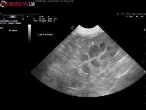
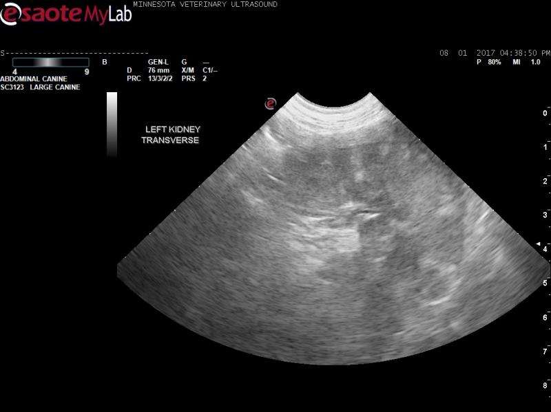
- Abdominal ultrasound done as part of a met check shows a coarse, micronodular spleen with multiple faint, poorly defined hypoechoic nodules. It also shows bilateral renal cortical echogenic densisities or small nodules.
- My differential list for the spleen includes benign hyperplasia vs. emerging hemangiosarcoma
- What is going on with the kidneys? Is this just cortical mineralization or could this perhaps be emerging neoplastic disease?


Comments
Causes of Focal increased
Causes of Focal increased cortical echogenicity:
-Wedge shaped chronic renal infarcts (not likely here)
-Primary or secondary neoplasia
-Infection?
-Parenchymal calcification
-Fibrosis
-Gas (unlikely here)
-Granulomas.
Probably would need a biopsy to sort it out. My most two likely considerations would be neoplasia vs granulomatous change
Thanks Randy. The owner was
Thanks Randy. The owner was considering splenectomy, so renal biopsies may be done at that time. Otherwise, I may never know. Appreciate your thoughts! I will bookmark them for next time.
-M
Renal infarcts are common
Renal infarcts are common with concurrent neoplasia in my experience and makes sense here since HSA is a vascular event disease. Also cortical mineralization orf the kidneys and spleen for that matter (side note) I see with emerging and full cushings cases… the sonographers calcinosis cutis:)