- 9 yr old 49 lb MN Pitbull presented for chronic, progressive dyspnea.
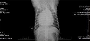
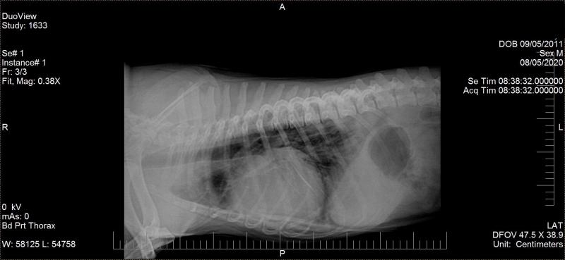
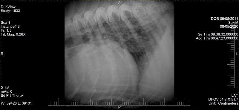
- Radiographs show cardiomegaly, pulmonary infiltrate, and possible pulmonary nodules
- HWT is antigen negative. Dog has not been on HW preventative.
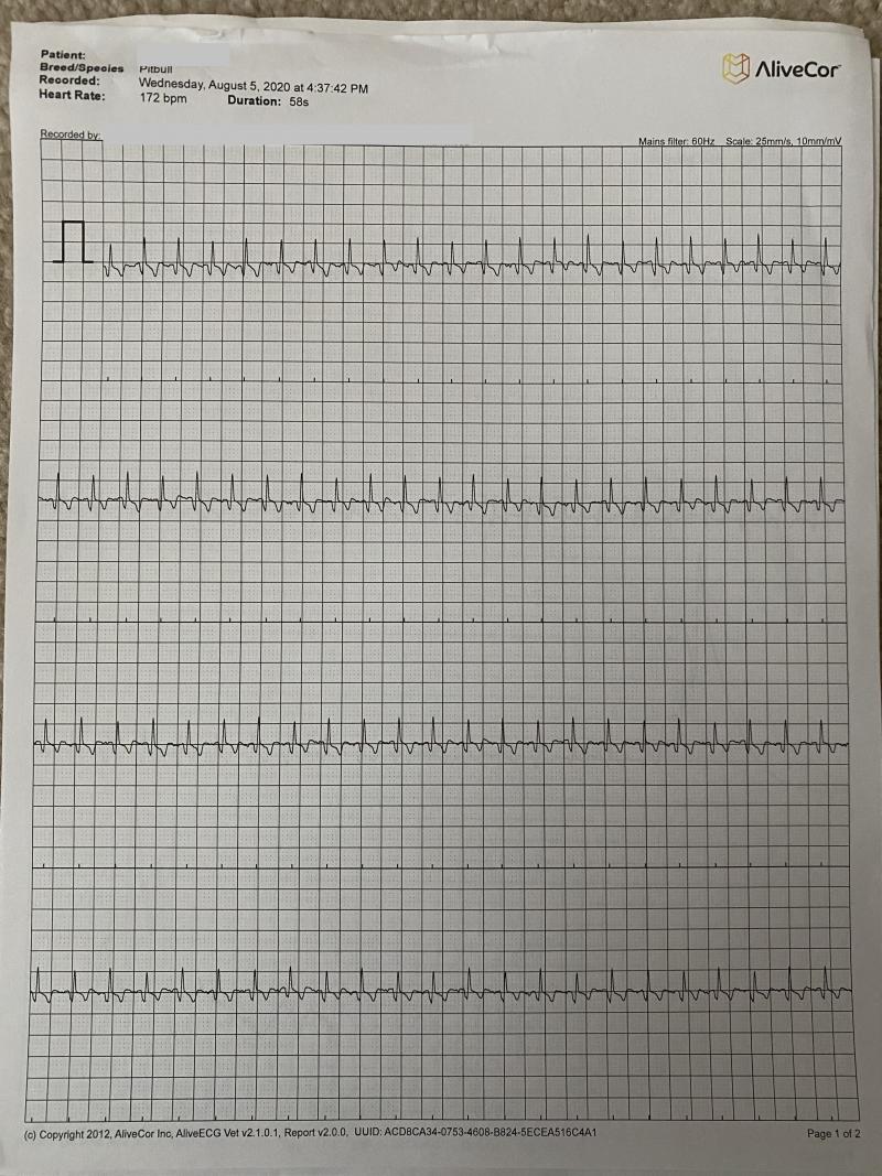
- ECG shows a regular sinus tachycardia
- Primary DVM reported that dog became cyanotic and collapsed when placed in lateral restraint for shaving. Lasix was administered about 15 minutes prior to echo.
- Brief echo performed with dog is sitting position. Dyspnea (marked chest wall movement and lung interference) interfered with Doppler studies.
- I see eccentric RV enlargement, MPA enlargement, flattening of the IVS on trans LV views. The LA is decreased in size.
- Although I don’t appreciate much if any RV concentric hypertrophy, I am concerned about pulmonary hypertension secondary to primary pulmonary disease (fungal, neoplasia, HW). Pulmonary emboli also on the list. I assume this is not right arrhythmogenic cardiomyopathy as an ECG performed twice showed no PVC’s. HR is elevated at 15-180bpm. LV measurements are normal and FS is high normal at 45%. I know TVI can cause RV enlargement, however, this dog has no prior hx of a murmur and decreased LA size would suggest that endocardiosis is not the primary disease. Again, would have loved to do Doppler but could not get still enough movement for a decent clip.
- Reccomended performing full bloodwork, microfilarial testing, and FNA of the lungs. Fungal titers also suggested but turn around time is slow. Medical management with antibiotics, bronchodilators, oxygen? Would like to do full echo with Doppler when dog is more stable.
- What are your thoughts?


Comments
yes big right heart without
yes big right heart without the eccentric hypertrophy I would think its an acute onset as the RV hasnt had time to muscle up and “train” for that increase in pulm pressure so its trying to “train fast” before disaster owing to a rapid pulm pressure increase … if that makes sense.
Pulmonary thromboembolism is first on my list when i see a sudden onset like this… Given the chronic respiratory but sudden acute onset, PTE secondary to chronic respiratory disease and other factors would be my concern… acute PTE on chronic respiratory. Maybe HW in the mix but I dont see any here. Ventricular septum is flattening and LA is normal so this is all right side.
keep grabbing rads on this guy you may see progresisve lung densities develoip over hours.
Sildenafil for sure to help the Pulm HT but the main issue is in the pulmonary tree. CT ideal of the chest.
Thank you!
Thank you!
Can also run a CRP looking
Can also run a CRP looking for inflammatory diseases. If fungal disease is presence, antibiotics can potentially worsen the outcome. As Eric mention, CT ideal and if doing it would also recommend bronchoscopy with BAL under the same anesthestic.
Ok, thank you Dr. Lobetti.
Ok, thank you Dr. Lobetti.