Hi everyone! just wanted to know if anyone has seen this type of vasculature before and whether there can be a normal variation to this? Apologies for the long history.
Minka is a 10 y.o FS American bulldog that was presented for weight loss, poor appetite, panting, pacing and restless behaviour. Owner reports of being unsettled with 3 occasions of smelly urine and lower back discomfort.
Previous medical history includes a grade 2 soft tissue sarcoma on the right hind limb which was removed on the 20/6/18. Surgery was uneventful. Histopathology reported wide margins suggestive of cure.
She is currently on Thyroxine supplement for hypothyroidism and her T4 was within normal limits of 22 nmol/L.
LDDS test revealed elevated resting cortisol of 156 nmol/L, post 3-hours of 161 nmol/L and post 8-hours of 153 nmol/L. (Normal 28-150nmol/L)
Blood work revealed elevation in liver enzymes ALP 231 IU/L, ALT 119 IU/L and hypocholesterolaemia 1.4 mmol/L.
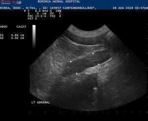
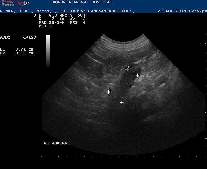
Both adrenal glands were enlarged;the left adrenal gland measured 0.86 cm in the caudal pole and 0.92 cm in the cranial pole. The right adrenal gland measured 0.71 cm in the caudal pole and 0.98 cm in the cranial pole. That being said, this dog does not look like a cushingoid dog to me. (i.e pot belly, alopeica, etc..) Bladder was fine.
There was an abnormally dilated vessel with the presence of turbulence in its lumen located in the right cranial abdomen. This vessel was observed curving around the right kidney cranially and dorsally. The vessel does not appear to be associated with the liver but was seen travelling near the liver before it curved back around towards the caudal abdomen.
Both the renal vein was also dilated and seen to be draining into this large dilated vessel suggestive of the caudal vena cava. However, it was not located in the correct position.
In addition, the caudal vena cava could be traced from the level of the aortic bifurcation cranially to the level of the right adrenal where it became difficult to trace.
At the level of the porta hepatis, it was also difficult to ascertain which is the portal vein as there were several dilated vessels around that region.
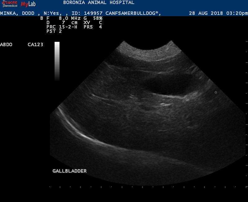
The liver didn’t look cirrhotic and the hepatic vessel was subjectively adequate. I’m not convinced the dog is cushingoid but not confident it has a shunt either. Any thoughts??


Comments
Thats a cool one looks like a
Thats a cool one looks like a rare portorenal or portocolonic extrahepatic shunt. Portorenal given where it ended up in the renal vein asd you describe. This is surely a “What’s that thing?” scan lol. Older dog shunt usually incidental in my experience. PDH looking adrenals I agree sometimes they plump first and become clinical later. Check BP & check a full cns exam to ensure no expaniove macroadenoma. I think people forget the CNS exam on PDH cushings patients often as macroadenomas go missed often the more we are doing CT at SonoPath we catch them.
Hi Eric, thanks for the
Hi Eric, thanks for the reply. As you can tell, this is the first time seeing a shunt like this and I can’t seem to find much information about it.
You mention it is an incidental in older dog? So the clinical signs should not be caused by the shunt then? Why/how does it happen? Does the renal vein bypass the CVC altogether and ends up in the Portal vein?
Although the LDDST is
Although the LDDST is indicative of Cushing’s disease, the clinical signs don’t fit. With the pacing and restless behaviour would look at primary neurological disease. Shunt is most likley a completely incidental finding.
Not sure about the exact
Not sure about the exact hemodyna,mics of the shunt since its so rare but just like azygois shunts that are seen in older animals despite being a congenital anomaly it all depends ont he shunt fraction, or the amount of blood shunted away from the portal system and away form the normal 75% of blood going to be metabolized by the liver through the portal system. Also depends ont he current hepatic integrity to continue to function enough to avoid clinical manifestations.
Gastrocaval shunts for example have a very high shunt fraction and manifest early in life typically… next in line splenocavals , then gastroazygos and then splenoazygos and so forth and the clinical shunt manifestations tend to be later in life on th eend of this list… not an exact science just a tendency in my experience. Anyone else out there share this tendency in shunt experience?
Could do a bile acid profile or ammonia level here and a full CNS exam +/- CNS CT with contrast.