Hello,
I can use some help with this case . Zoey is a 7 years old lab X that presented for ascites
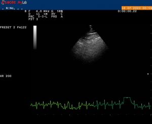
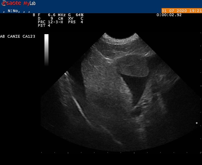
U/S : no macroscopic neoplasia, liver was subjectivelly small but did not looked cyrrhotic to me ( My understanding is that U/S can underestimate early cirrhotic appearance) . No hepatic congestion, no distended CBC. No pericardial effusion. No PLN. Albumin is low 16.9 before Prednisone trial then 19/L ( not low enough to explain ascites) Thoracic X-rays were normal
We’ve tried Prednisone at 2 mg/ kg daily and Spirinolactone as a treatment for PLE with no improvment after 2 weeks
Bile acids are both in high 80/88. I suspect cyrhosis with secondary Portal hypertension but not convinced . Did FNA of the liver, results are pending .
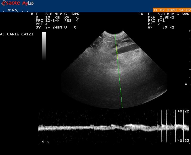
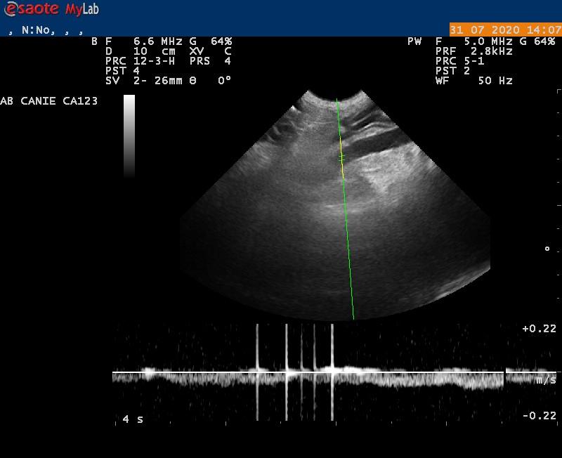
Does this look like liver cyrrhosis to you ? Does my PW help to confirm, rule out Portal Hypertension ?
Thank you !
Calin
Comments
Liver doesnt look bad enough
Liver doesnt look bad enough to me in fact only minor changes so likely not a player. PLE in large breeds always be concerned for lymphoma and seocndary PLE. Pred may be suppressing it now. You can cytospin the ascites and slide the sediment right away and see if anything says lymphoma. As you correctly say since albumin is not < 1.5 then hydrostatic issues are in play and usually thats a lymphatic congestion issue.
My “Working thorugh ascites” by means of the probe lecture may help here. Please excuse the shameless plug:)
scroll down on this link
https://www.shopsonopath.com/online-ultrasound-courses
See FNA liver attached. Would
See FNA liver attached. Would you still suspect PLE/ Lymphoma and consequently treat for it of still chase the liver with a biopsy ?
WE are getting to the end of the clients resources and I m contemplating treating liver cirrhosis + tx for PLE …???
Thank you !
Again on the single still
Again on the single still image of the liver it doesnt look that bad. Usually portal hypertension cases will have significant diffuse disease on the sonogram as the liver is a very resilient organ and can handle insults and not fail or cause portal hypertension til end stage. I would also expect bun to drop along with albumin and then glucose. Your pv velocity is off line and not < 15 deg in line with the portal flow so its not a valid number and pv velocity is very very unreliable in general as I’ve seen end stage liver with portal hypertension with normal pv velocities and low velocities in relatively normal liver so honestly i just stopped doing them despite proper lineup but thats me:) With portal hypertension the pancreas should be edematous as well and often secondray shunting cranial medial to the kidneys and more often than not splenic congestion but thats variable. Bile acids elevation can also occur with PLE/intestinal dysbiosis so not necessaruly liver based. Fibrosis cannot be assumed based on an fna that doesnt exfoliate. Needs a core bx. I have seen fulminant peracute liver failure cases do this however but very rarely. I dont personally value fluid samples if not cytospun immediately and sediment evaluated for cell dx. The fluid description unfortunately doesnt add anything as it could be lymphomatosis suppressed by the pred. Still a mystery here unfortunately.
Thank you EL.
Will
Thank you EL.
Will concentrate our treatments toward managing ascites from PLE.