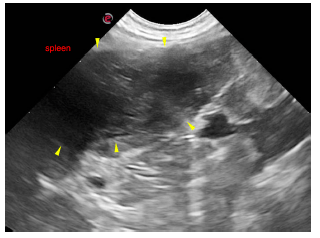
This 9 year old FS dog presented with a one month history of weight loss, ADR. Splenic sample submitted for telecytology too.
Physical Exam: Thin, palpable abdominal left sided mass
CBC/Chem: ALT 170
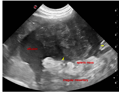
This 9 year old FS dog presented with a one month history of weight loss, ADR. Splenic sample submitted for telecytology too.
Physical Exam: Thin, palpable abdominal left sided mass
CBC/Chem: ALT 170