Case Study
RADS – Congestive Heart Failure and Pulmonary Edema, possible Liver Neoplasia vs Cushing’s in a 12 year old FS dog
Image Interpretation
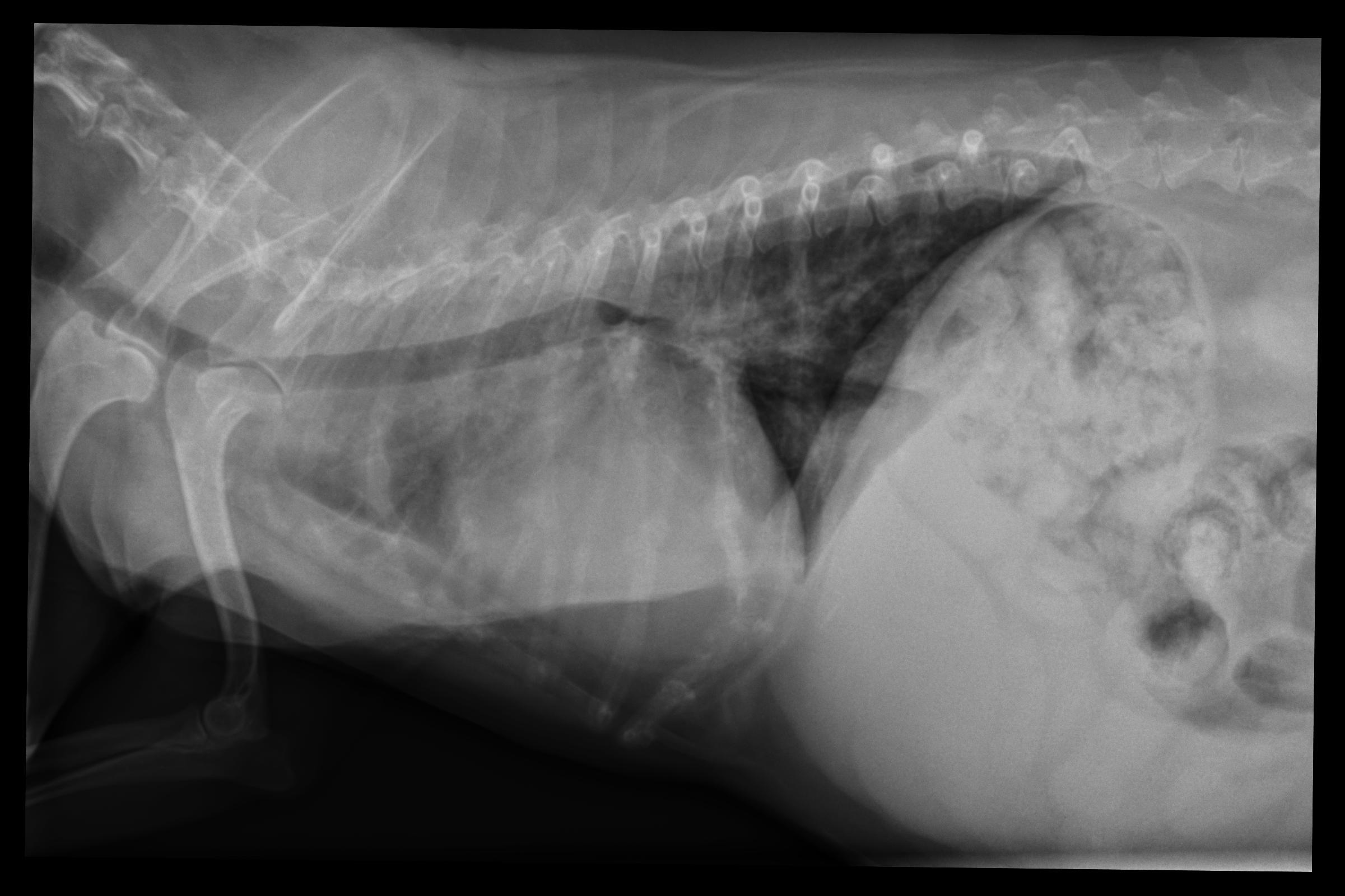
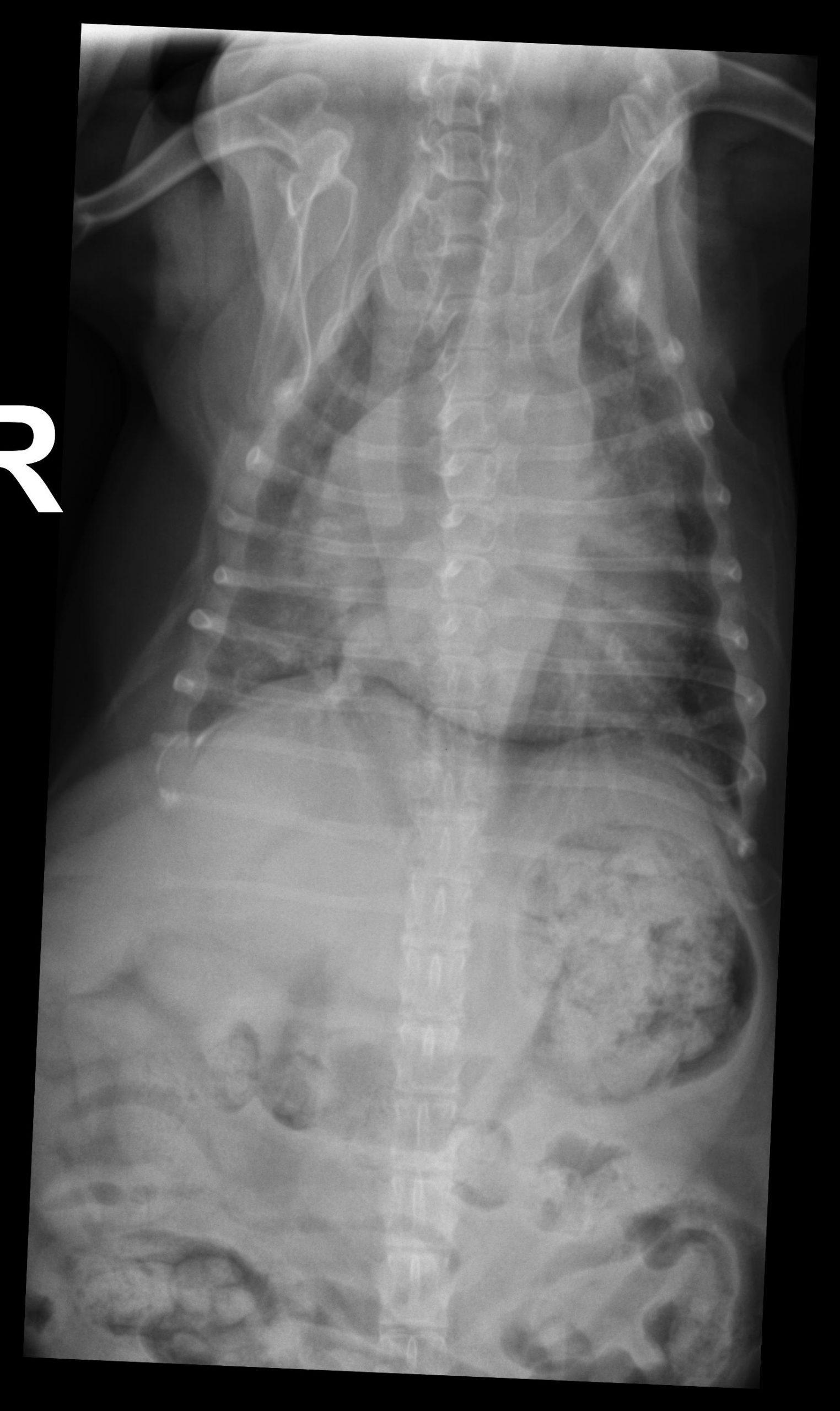
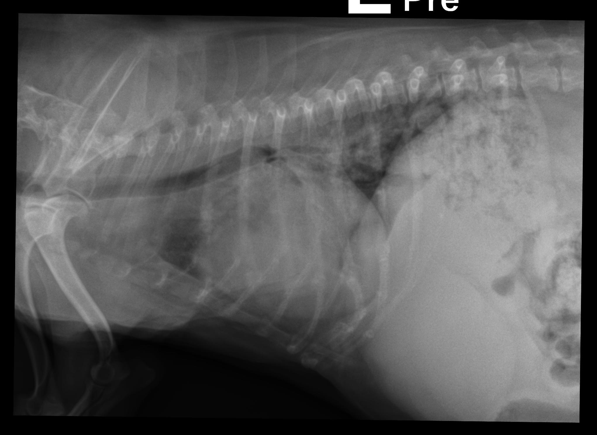
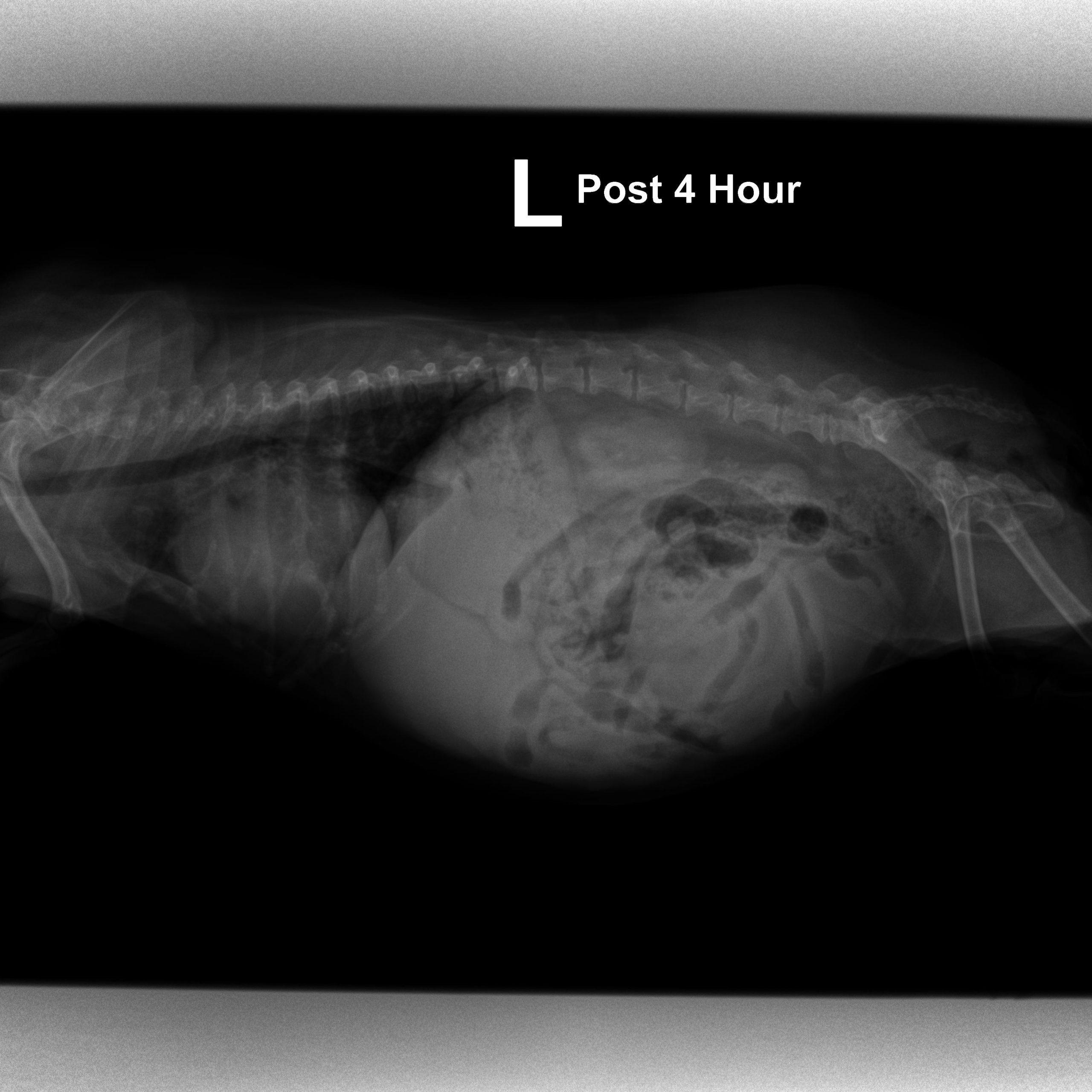
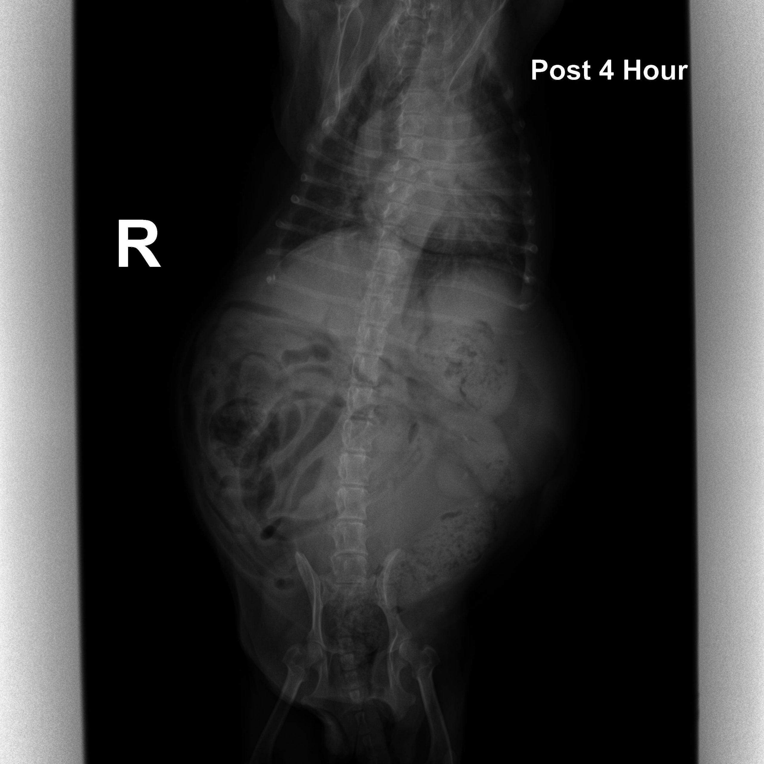
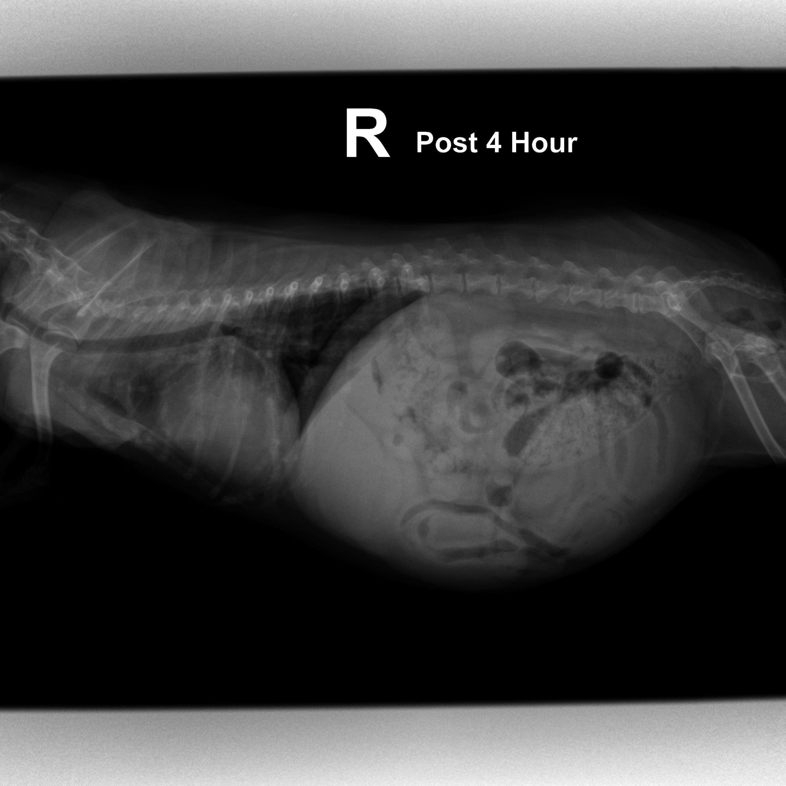
left lateral and DV thorax, baseline and 4 hours post diuretics: The patient was potbellied and obese. The chest volume was small, the abdominal volume was large. The abdominal wall was pendulous.
Osseous structures: Overall moderate degenerative changes were associated with the axial skeleton. There was moderate generalized osteopenia.
Intrathoracic structures: The esophagus contained a mild amount of gas.
Abdominal views:The abdominal serosal detail was influenced by crowding of the abdominal organs. The liver presented severe asymmetrical enlargement.
The stomach was elevated.
The splenic tail was not well delineated against the enlarged liver.
Multifocal mineral opacities were associated with the region of the kidneys and adrenal glands.
The urinary bladder was severely distended.
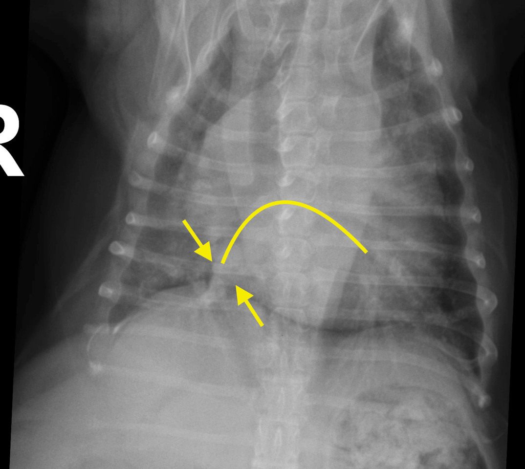
The trachea was elevated.
The cardiac silhouette was enlarged with loss of the caudal cardiac waist and split main stem bronchi.
The pulmonary veins were dilated on the initial radiographs. On the control radiographs the venous congestion was largely resolved.
There was a mixed alveolar-interstitial pulmonary infiltrate emphasizing the perihilar region on the initial radiographs which had resolved after 4 hours.
DX
Comments
|
There were signs suggestive for bilateral renal calculi which require clarification against dystrophic/carcinomatous adrenal gland calcification by means of abdominal ultrasound. Next to abdominal ultrasound further diagnostic workup requires a full cardiac echo. |
Patient Information
Images