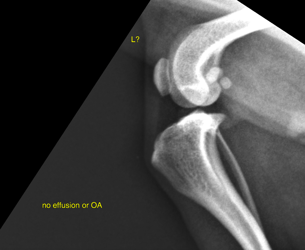
This 7 year old F Chihuahua dog presented with acute left hind leg lameness of 2 days duration. Pain on extension of left stifle, positive cranial drawer.
This 7 year old F Chihuahua dog presented with acute left hind leg lameness of 2 days duration. Pain on extension of left stifle, positive cranial drawer.