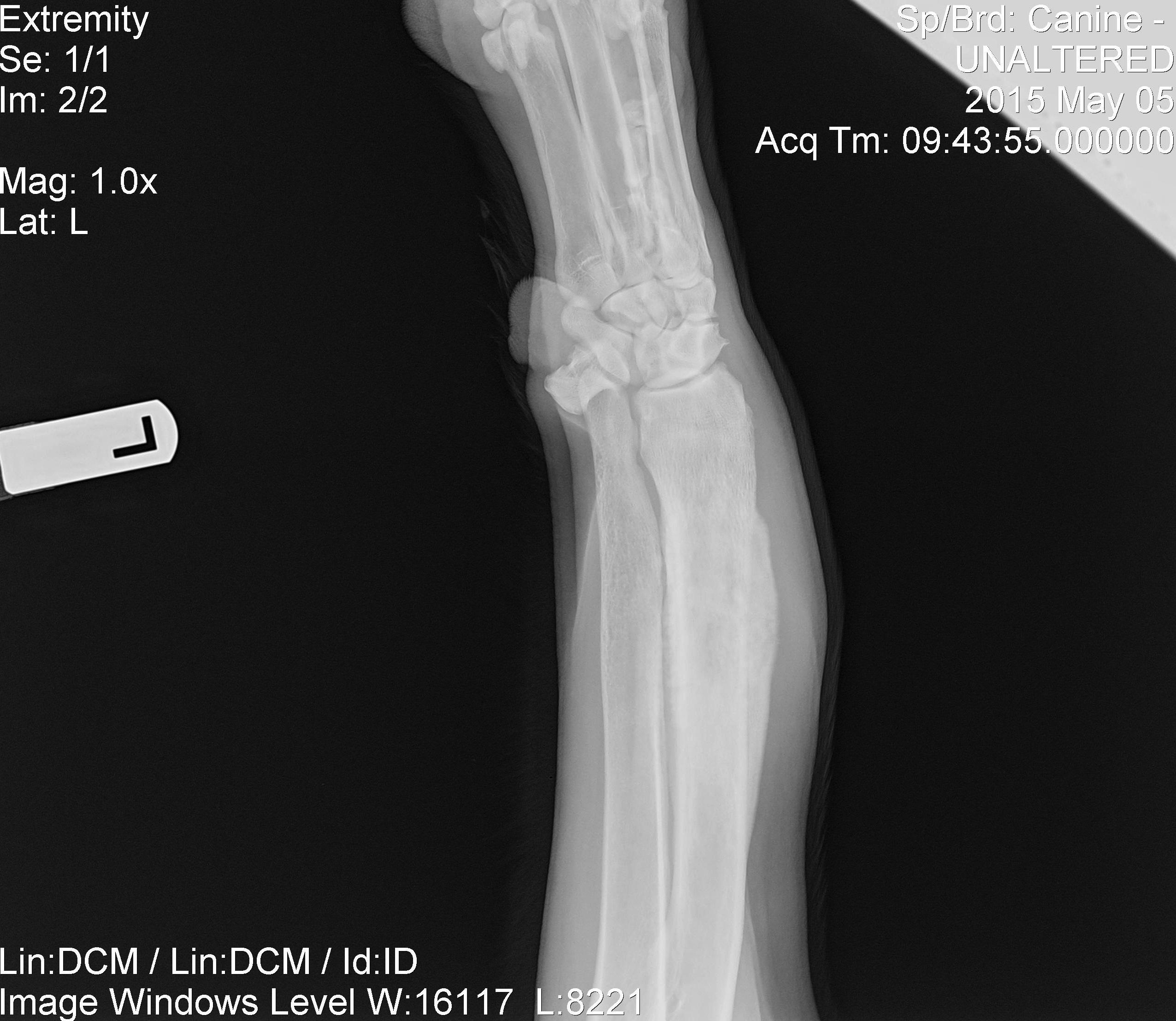
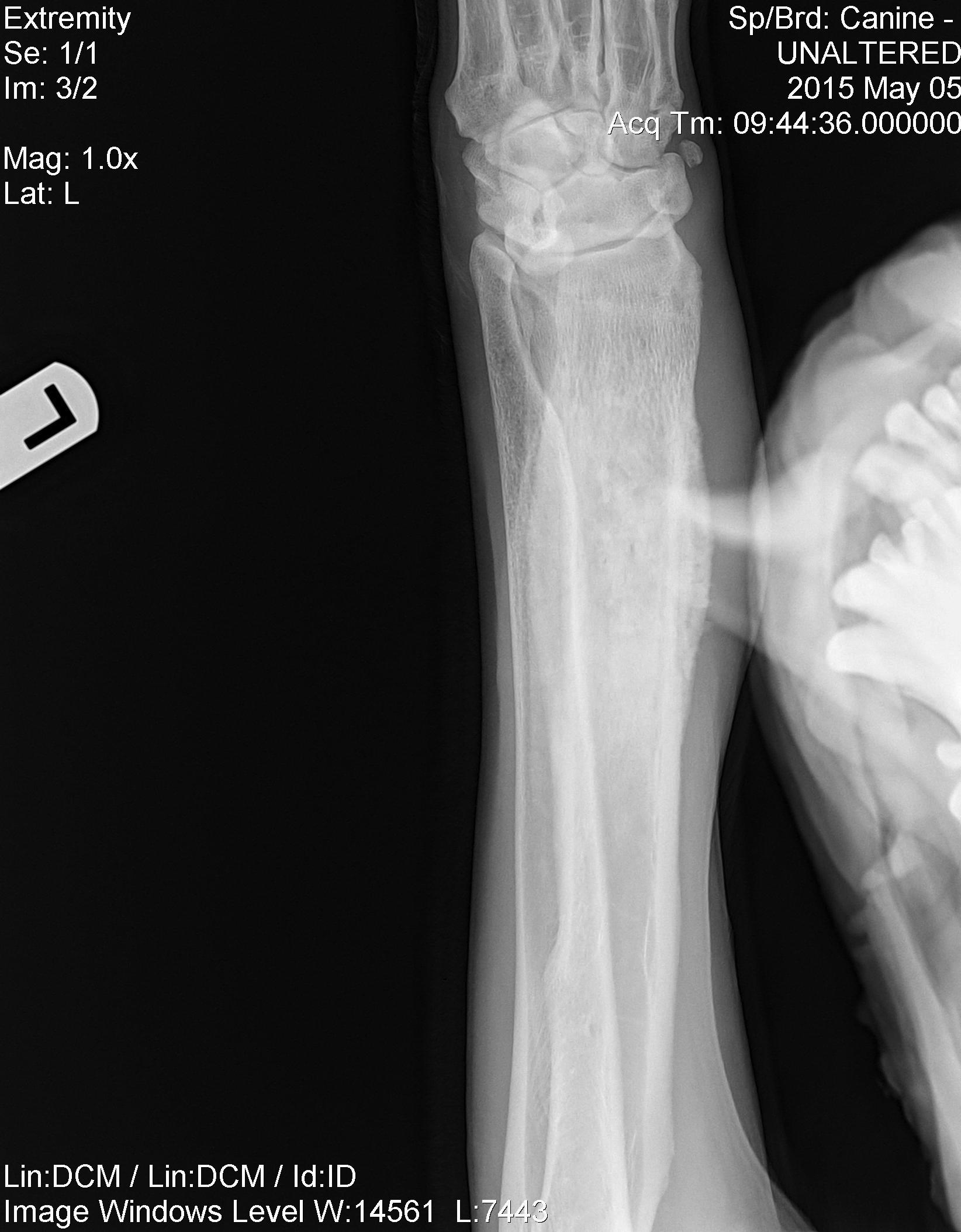
A 9-year-old MN K9 was presented for lameness of the right front leg.
A 9-year-old MN K9 was presented for lameness of the right front leg.
A 9-year-old MN K9 was presented for lameness of the right front leg.
A 9-year-old MN K9 was presented for lameness of the right front leg.
Right Humerus: A 6 hole metallic plate is noted extending from the proximal metaphyseal region to the distal metaphyseal region of the right humerus. The implant in the proximal bone fragment are well aligned and appear intact. The screws within the distal fragment appear to not be in contact with the cortical bone. The plate is displaced caudally and laterally. This is resulting in a severe malalignment (with override and severe angular malformation) of the long oblique distal diaphyseal humeral fracture. The wires are distracted. There is soft tissue swelling at the fracture site.
Right Elbow: Moderate degenerative joint disease. Discontinuity of the contour of the medial coronary process and a step between the medial coronoid and the medial humeral condyle. The anconeal process is not seen while on this extended lateral projection.
The patient was recommended for the following: 1. Considering the patient’s clinical signs, consider stabilization of the humeral fracture. This will require an open reduction. However, the open reduction may be limited by the soft tissue component to the healing process, which is not well appreciated on these images. 2. Consider further work-up of the elbow joint for the underlying degenerative process. This may not be possible, if the cause was trauma associated with the humeral fracture