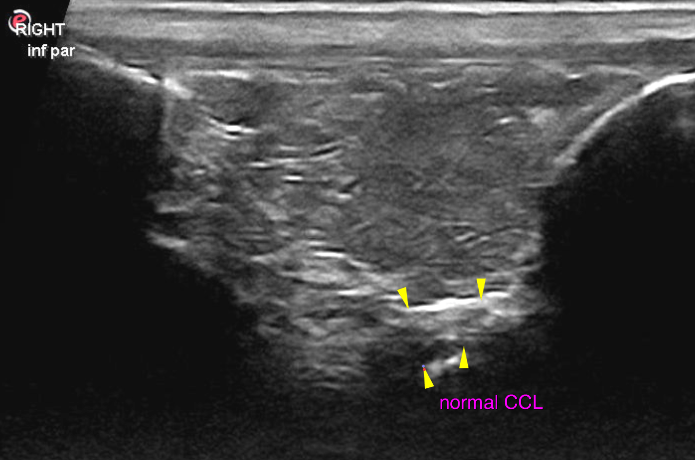
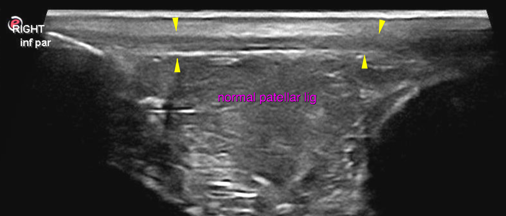
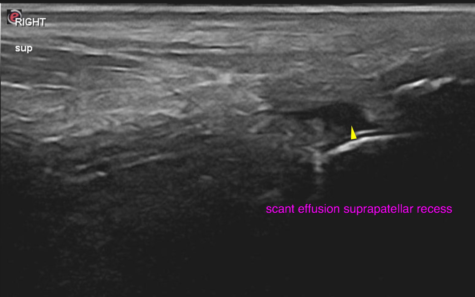
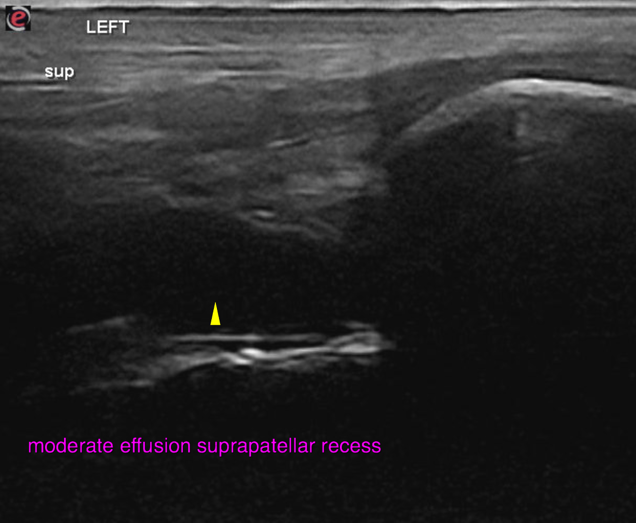
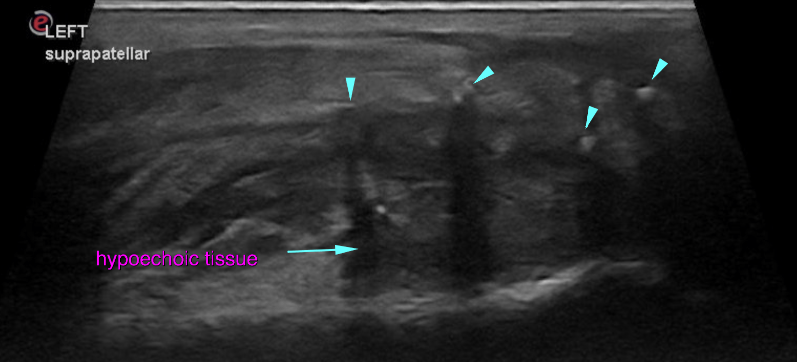
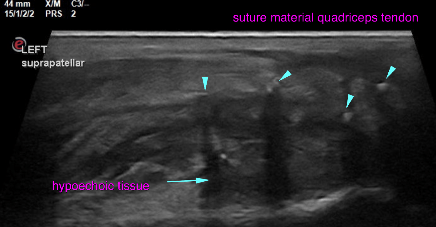
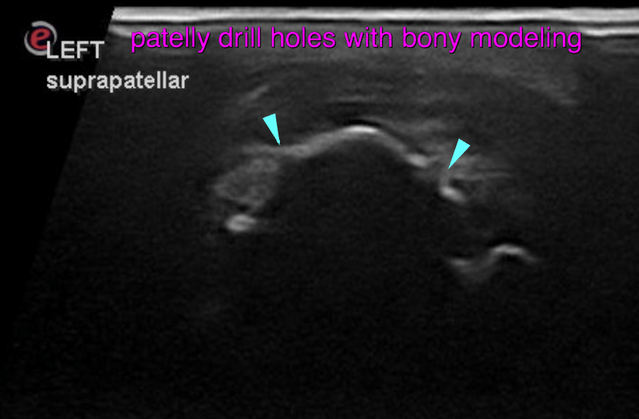
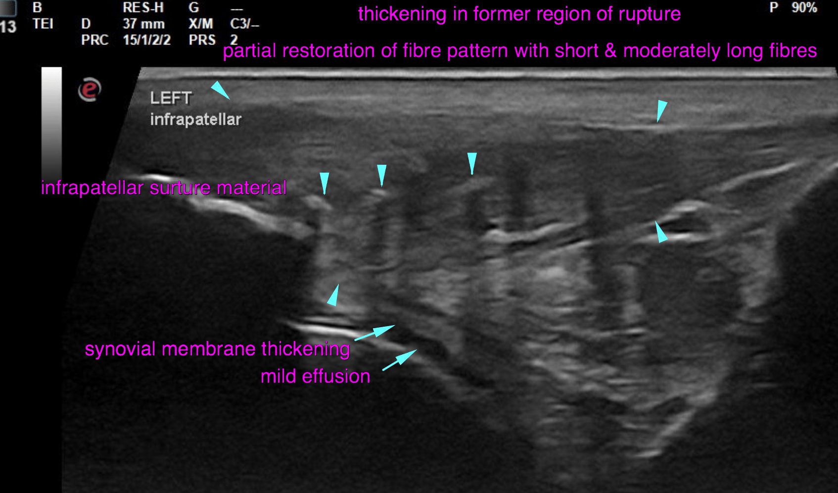
This 9 month old M intact German Wirehaired Pointer dog presented with left hind lameness of several months duration. Suspect traumatic injury to patellar ligament.
This 9 month old M intact German Wirehaired Pointer dog presented with left hind lameness of several months duration. Suspect traumatic injury to patellar ligament.
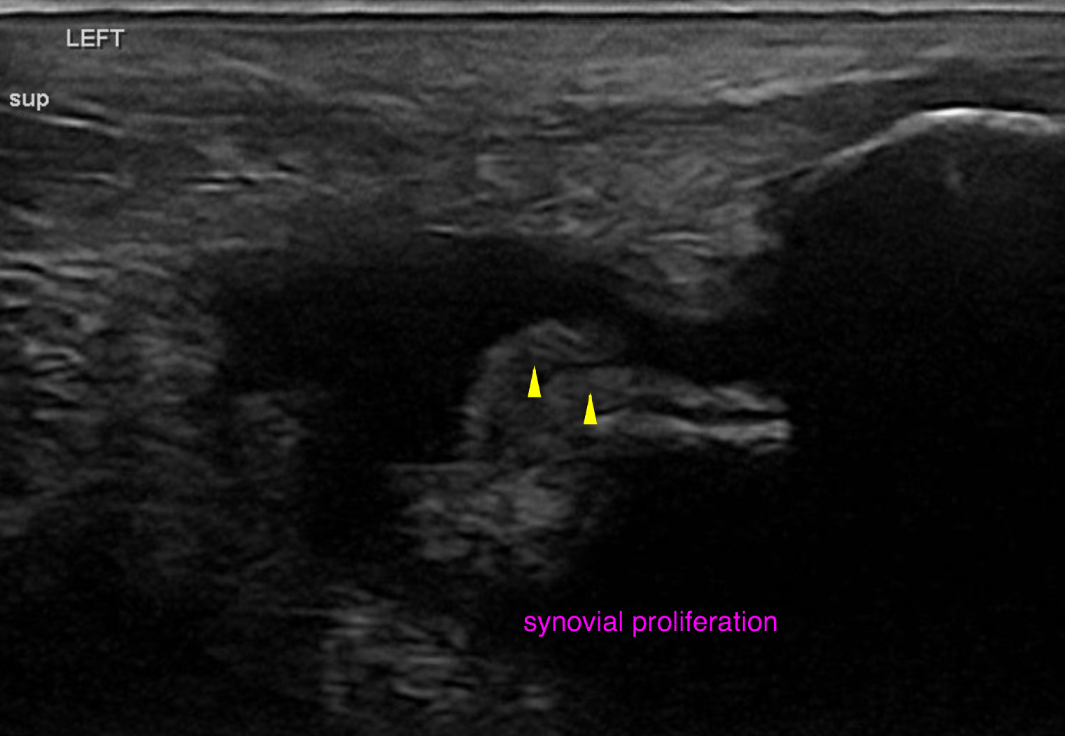
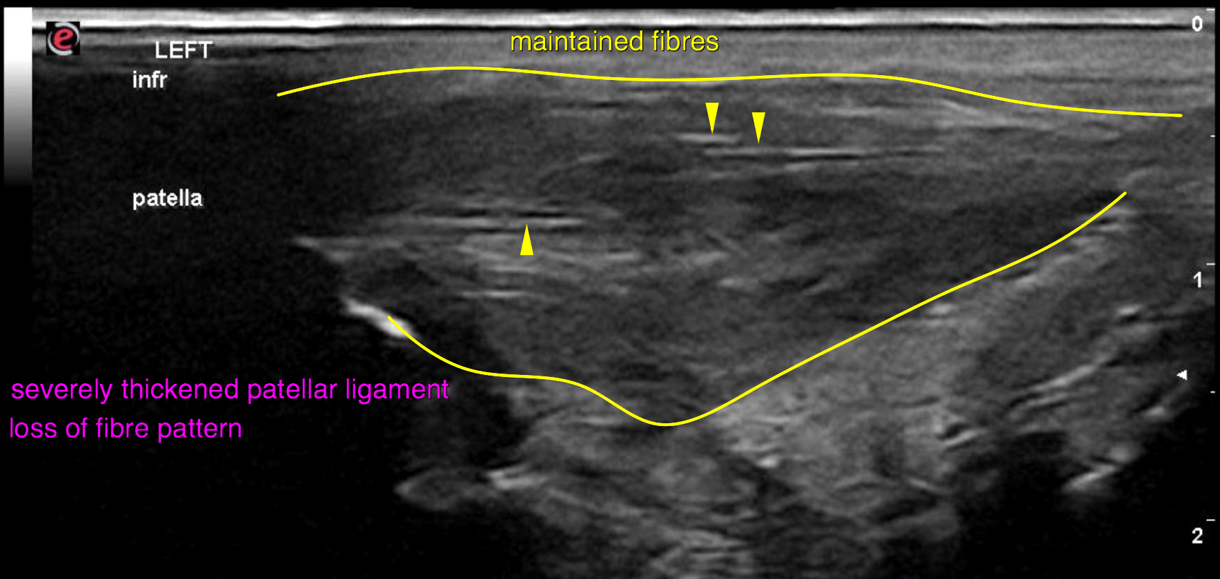
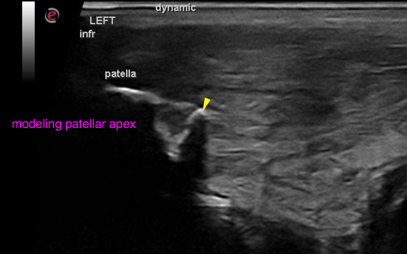
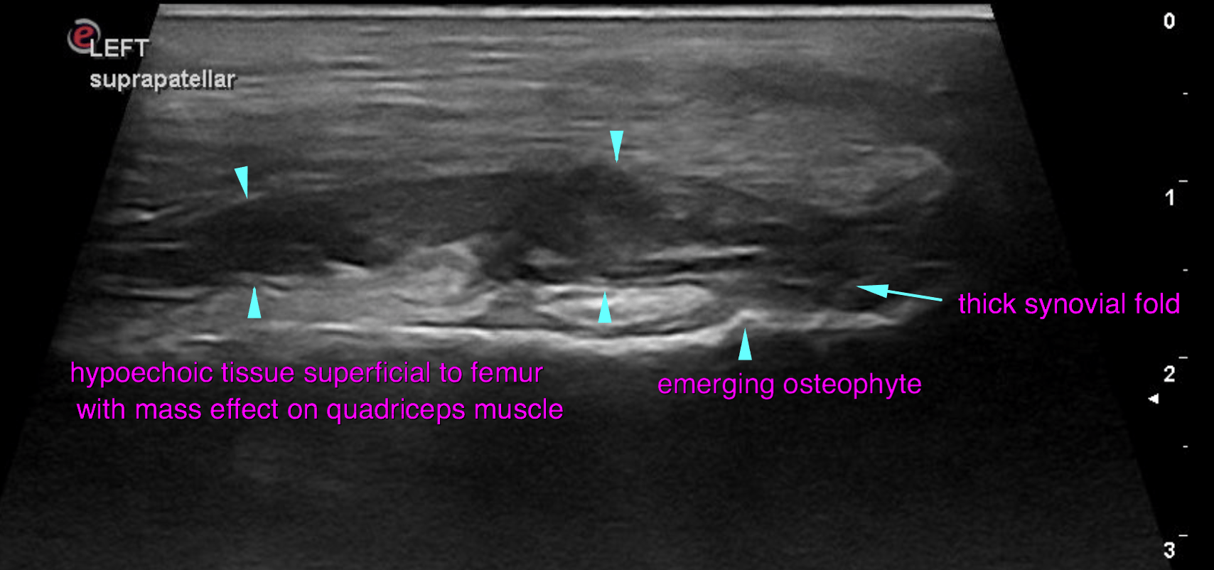
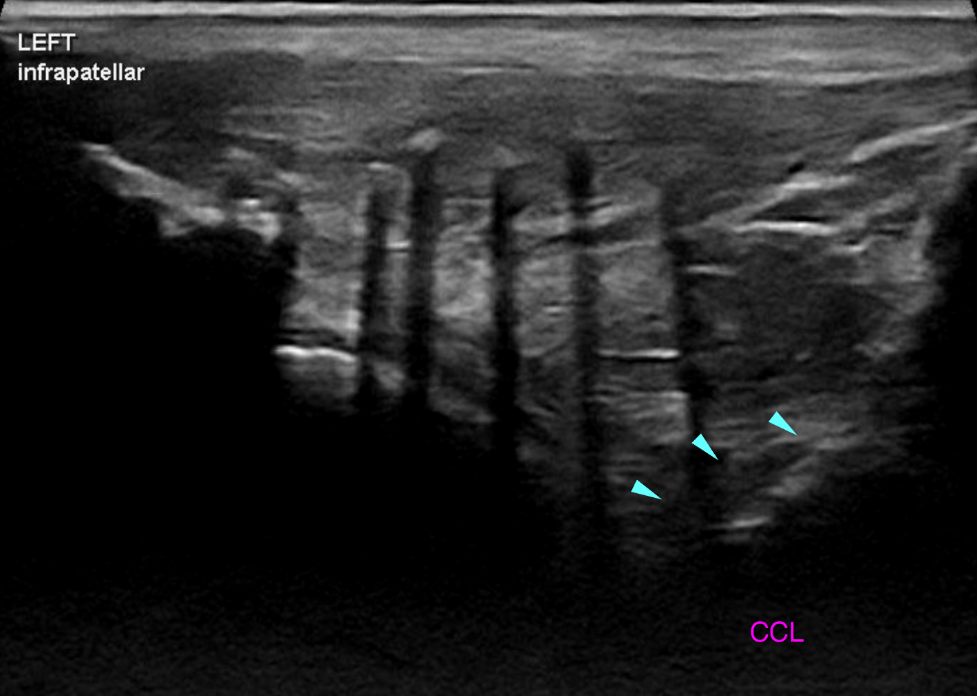
Physical exam: Gait/Lameness- Grade III/V left pelvic limb lameness Posture: Off weighting left pelvic limb at a stand. Joint palpation left stifle – Severe patellar ligament swelling, pain on extension of stifle, no cruciate instability, possible defect on the lateral aspect of the origin of the patellar ligament. Healed ~1cm diameter dermal scar over the proximal patellar ligament. Mild muscle atrophy of the left pelvic limb