Skip to content
Customize Consent Preferences

We use cookies to help you navigate efficiently and perform certain functions. You will find detailed information about all cookies under each consent category below.

The cookies that are categorized as "Necessary" are stored on your browser as they are essential for enabling the basic functionalities of the site. ... 

Always Active

Necessary cookies are required to enable the basic features of this site, such as providing secure log-in or adjusting your consent preferences. These cookies do not store any personally identifiable data.

No cookies to display.

Functional cookies help perform certain functionalities like sharing the content of the website on social media platforms, collecting feedback, and other third-party features.

No cookies to display.

Analytical cookies are used to understand how visitors interact with the website. These cookies help provide information on metrics such as the number of visitors, bounce rate, traffic source, etc.

No cookies to display.

Performance cookies are used to understand and analyze the key performance indexes of the website which helps in delivering a better user experience for the visitors.

No cookies to display.

Advertisement cookies are used to provide visitors with customized advertisements based on the pages you visited previously and to analyze the effectiveness of the ad campaigns.

No cookies to display.

ORTHO – Chronic degenerative joint disease (DJD) with synovitis, joint effusion and partial cranial cruciate ligament (CCL) rupture in a 2 year old F Labrador Retriever dog

Case Study

ORTHO – Chronic degenerative joint disease (DJD) with synovitis, joint effusion and partial cranial cruciate ligament (CCL) rupture in a 2 year old F Labrador Retriever dog

This 2 year old F Labrador Retriever dog presented with apparent left cruciate tear 3 month ago

This 2 year old F Labrador Retriever dog presented with apparent left cruciate tear 3 month ago

Image Interpretation

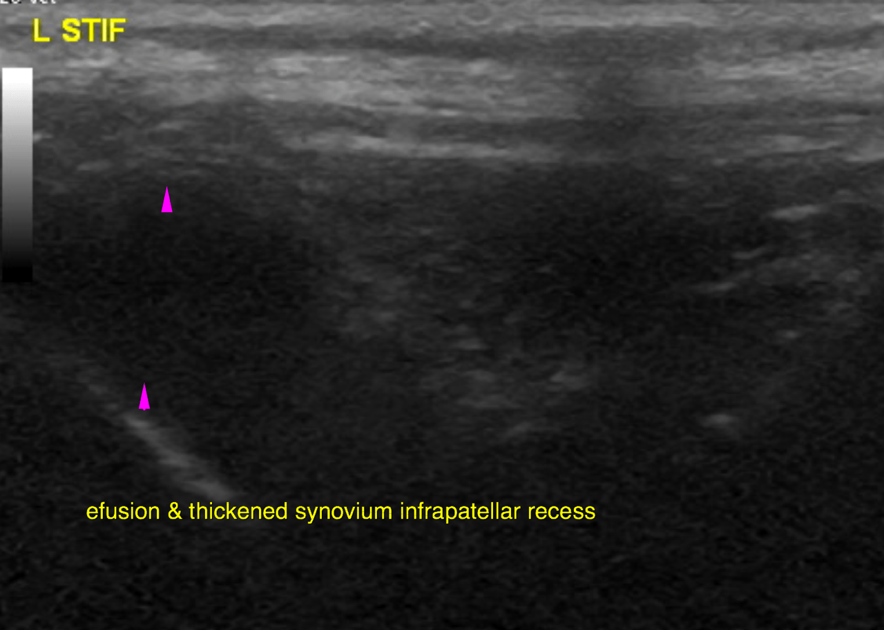
Ultrasound of the left stifle – Both the supra- and infrapatellar recess of the left stifle joint reveal mild synovial
thickening as well as mild anechoic effusion.
Emerging osteophytes are seen at the periarticular margins of the femoropatellar joint
(mainly at the proximal extent of the femoral trochlea).
The view on the cranial cruciate ligament (CCL) is limited. It appears to be continuous
and hypoechoic, but slightly ill defined and irregular in thickness. Scant
periligamentous effusion is seen.
The infrapatellar fat body presents mild heterogeneity as found commonly in
degenerative joint disease (DJD). 
The cranial and mid portion of the medial meniscus are well visible and in situ with
even surface and uniform echogenicity.

Right stifle – wnl

DX

The findings of the left stifle joint are compatible with mild generalized chronic degenerative joint disease (DJD) with synovitis and mild joint effusion as well as emerging osteophytosis. The findings of the cranial cruciate ligament suggest edema and partial/developing rupture. Progression to biomechanical failure has to be expected.

Outcome

There is no evidence of medial meniscal injury.
The ultrasonographic examination of the right stifle joint is within normal limits at
this point

Patient Information

Patient Name : Calli Parisi/North Penn
Gender : Female, Intact
Species : Canine
Type of Imaging : Ultrasound
Status : Complete
Liz Wuz Here : Yes
Code : 18_00007

Clinical Signs

  • Lameness

History

  • Cruciate ligament injury

Images

bildschirmfoto_2016-05-12_um_08bildschirmfoto_2016-05-12_um_09bildschirmfoto_2016-05-12_um_09bildschirmfoto_2016-05-12_um_09

Clinical Signs

  • Lameness