This 7 year old FS Labrador Retriever dog presented with left hind lameness and a swollen left hock
This 7 year old FS Labrador Retriever dog presented with left hind lameness and a swollen left hock
This 7 year old FS Labrador Retriever dog presented with left hind lameness and a swollen left hock
This 7 year old FS Labrador Retriever dog presented with left hind lameness and a swollen left hock
ultrasound of the achilles –
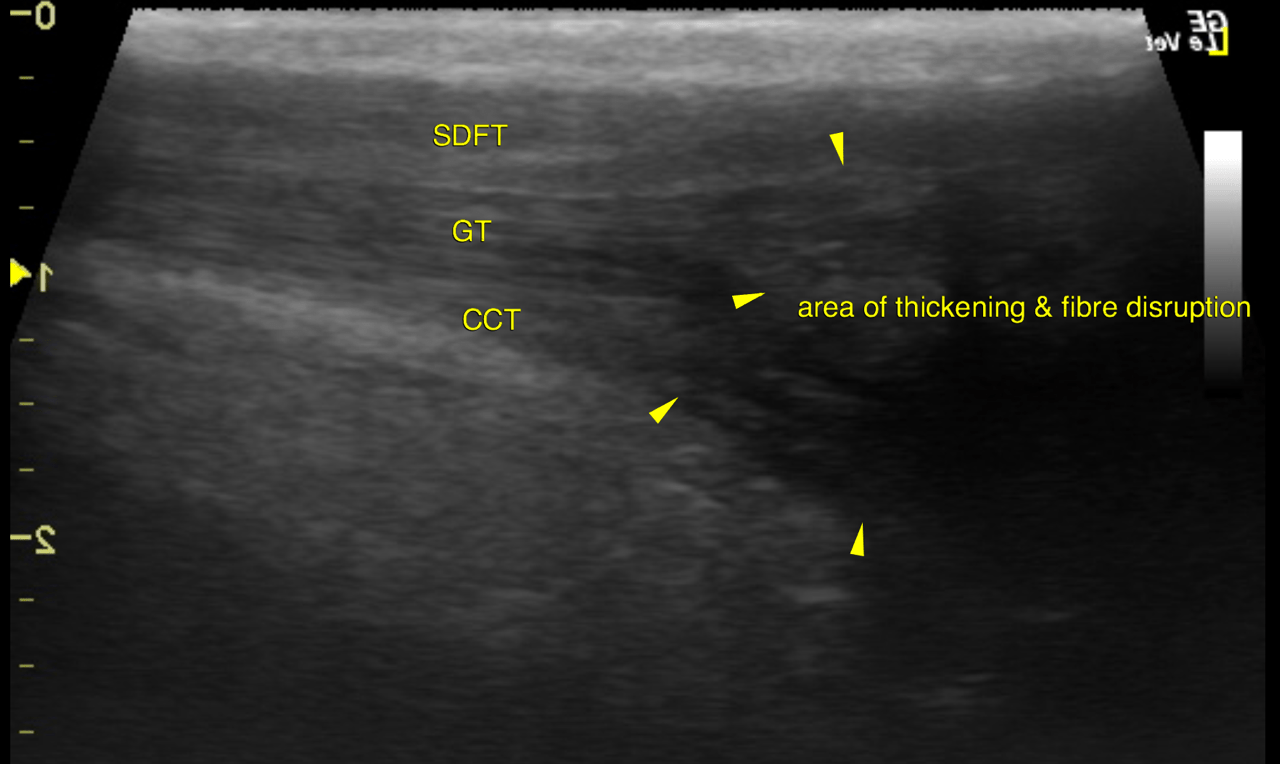
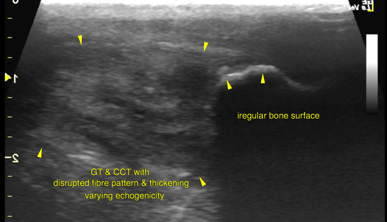
Left: Marked localized swelling with fibre disruption and varying degrees of echogenicity is noted for the distal gastrocnemius and common calcaneal tendons next to their calcaneal insertion. The affected region spans approximately 3 cm proximal to the calcaneal tubercle. The maximal diameter of the swelling is 2 cm. The bone surface of the calcaneal tubercle presents marked irregularity and modeling with concave defects. One larger and multiple small rounded bony bodies with echogenic surface and solid distal shadowing are seen within the affected region next to the calcaneal tubercle.
Right:
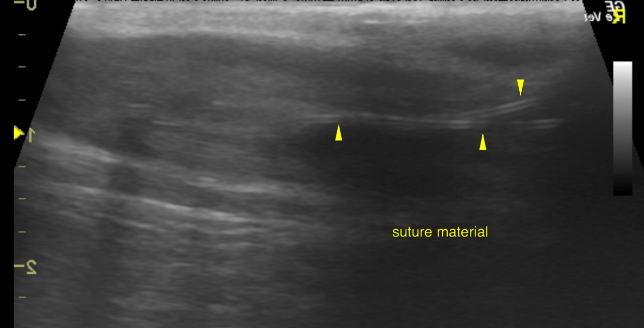
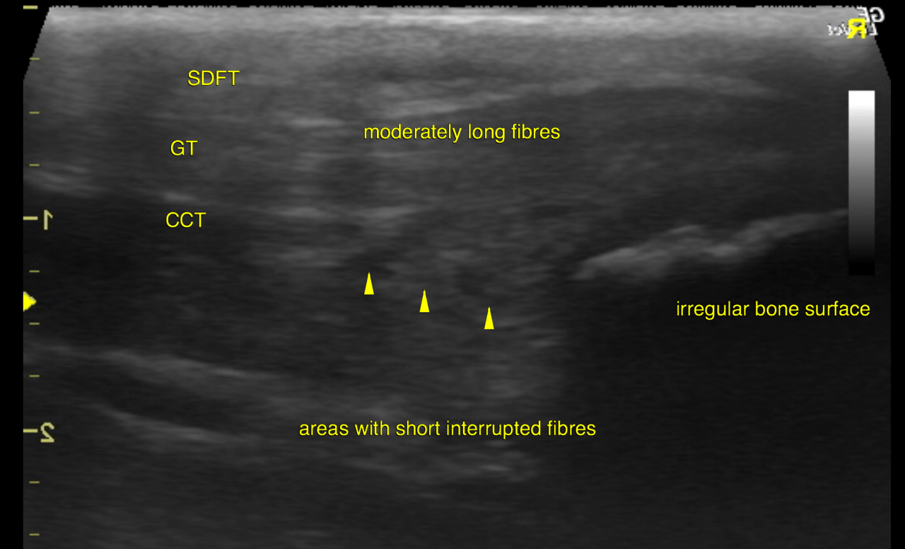
An elongated swelling of the distal gastrocnemius and common calcaneal tendons is seen up to their calcaneal insertion. The affected region of the gastrocnemius tendon reveals restoration of moderately long fibres of uniform echogenicity. The replacement tissue of the common calcaneal tendon presents shorter fibres with less uniform distribution. Long loops of suture material are seen within both components of the deep Achilles tendon. The bone surface of the calcaneal tubercle presents marked irregularity and modeling. One larger rounded bony body with echogenic surface and solid distal shadowing is seen next to the calcaneal tubercle.
left: Chronic degeneration with repetitive microruptures – such as in Doberman – appears to
be the most likely underlying pathology here especially since it is a bilateral disease.
The typical clinical presentation at the point of biomechanical failure is toe tipping
stance & gait.
At this advanced stage response to conservative management is assumed to be
inappropriate. Successful healing is likely to require temporary fixation with or without
tendon suture depending on the mode of fixation.
right: According to the ulrtrasonographic quality restoration of stability
appears to be appropriate for the gastrocnemius tendon and slightly lag behind the
anticipated degree of healing for the common calcaneal tendon.