Tnis 6 year old FS Labrador Retriever dog presented with right front lameness
Physical exam: tender right shoulder, swelling right carpus
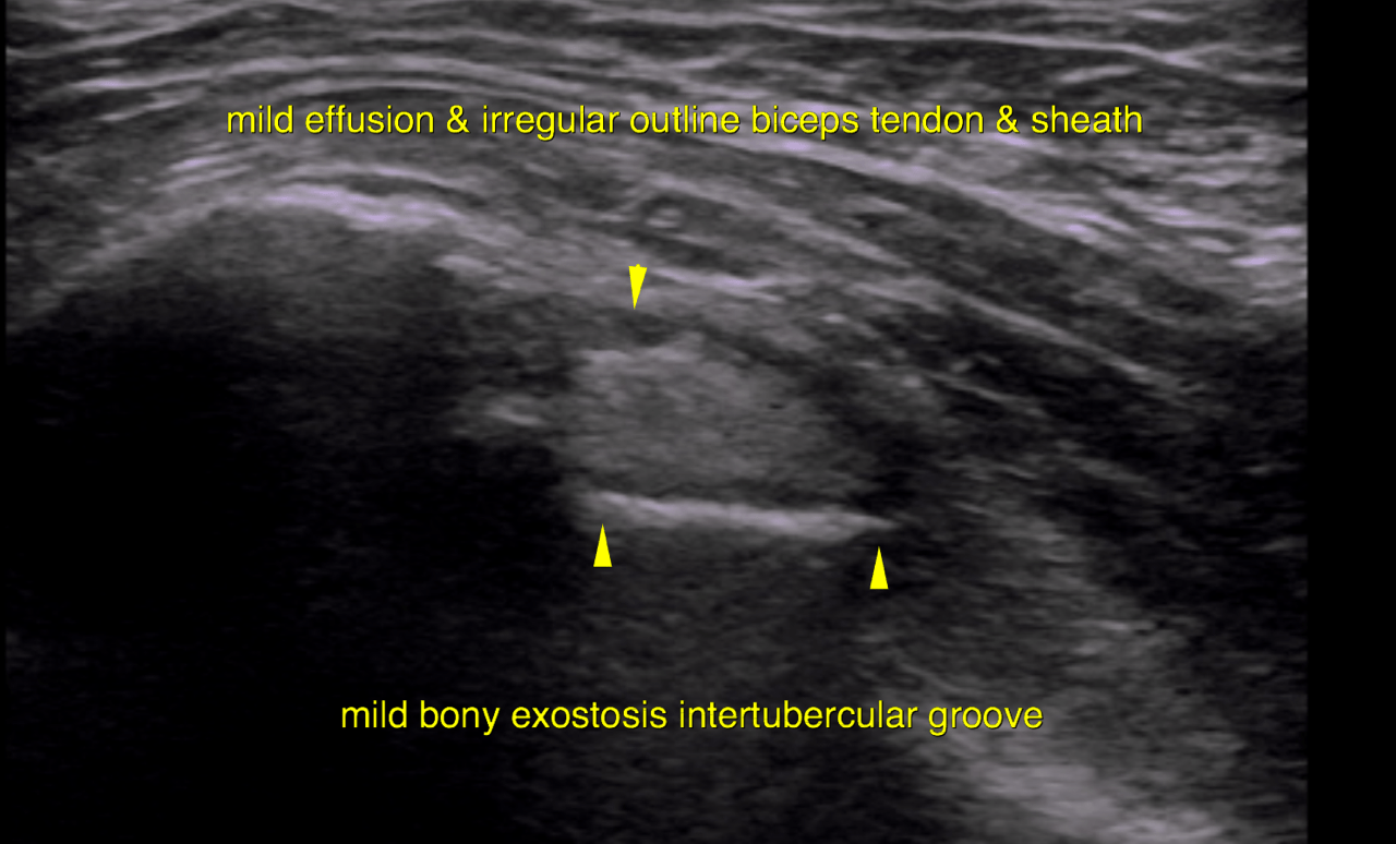
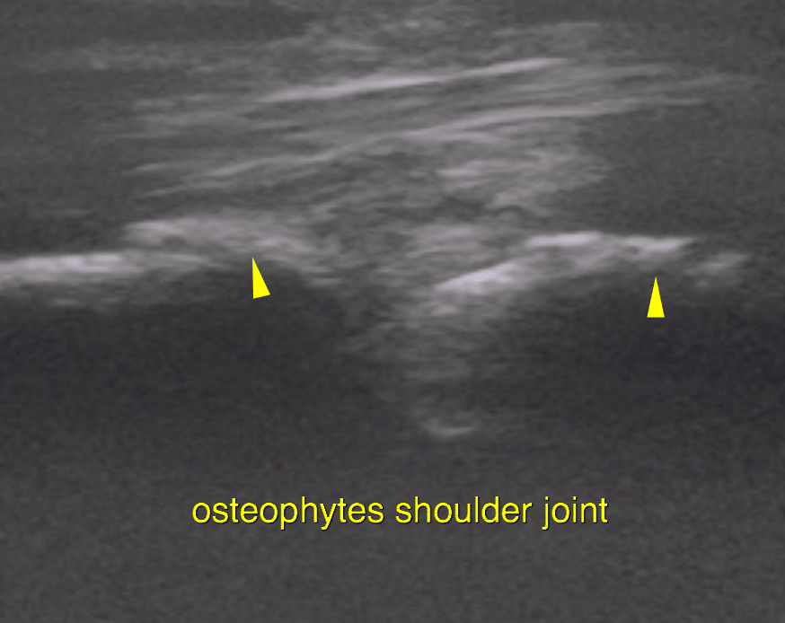
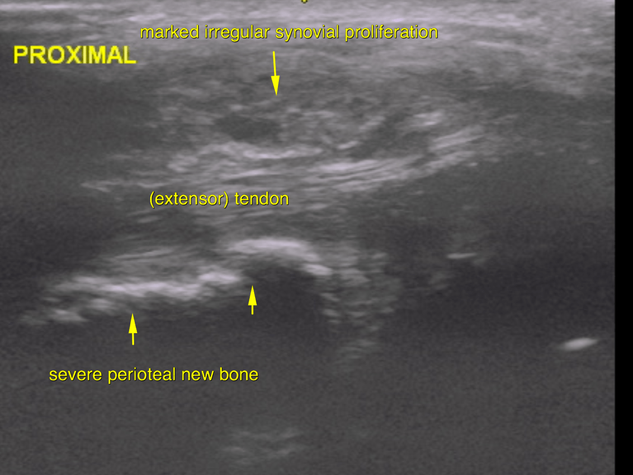
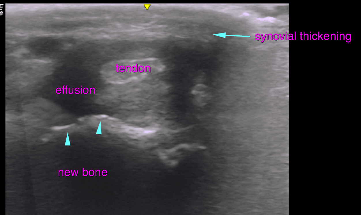
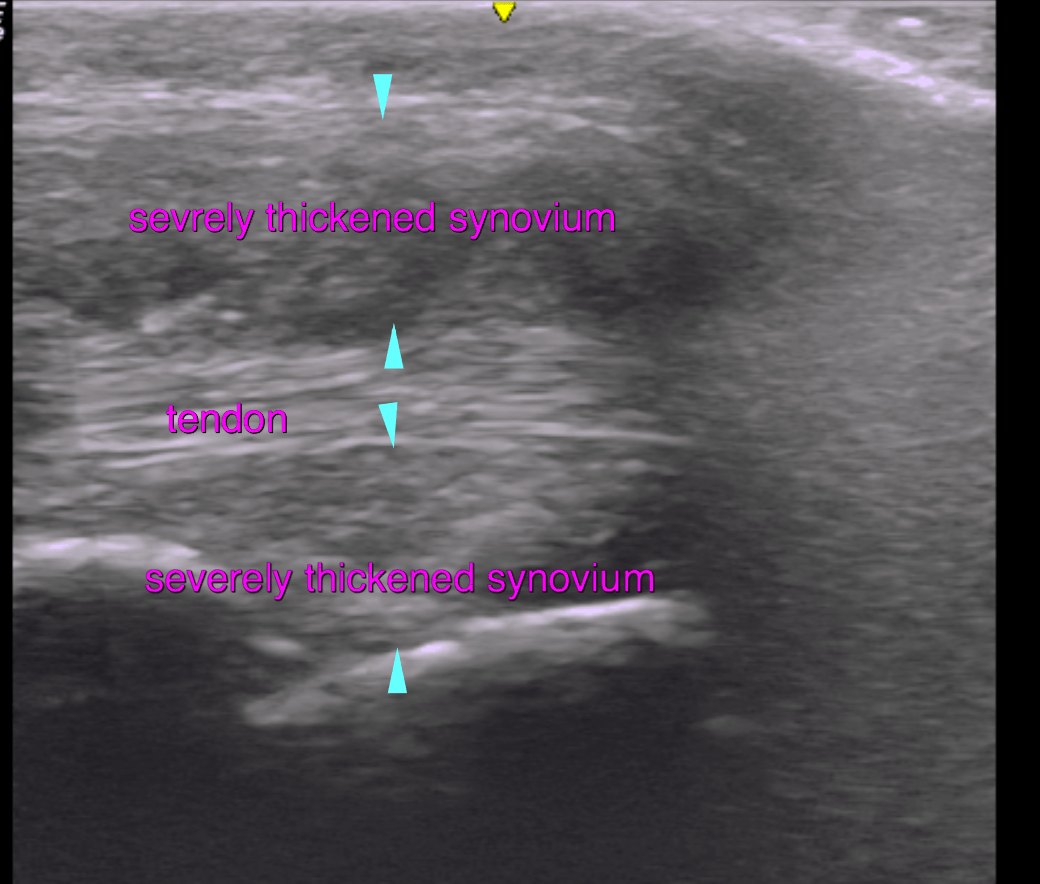
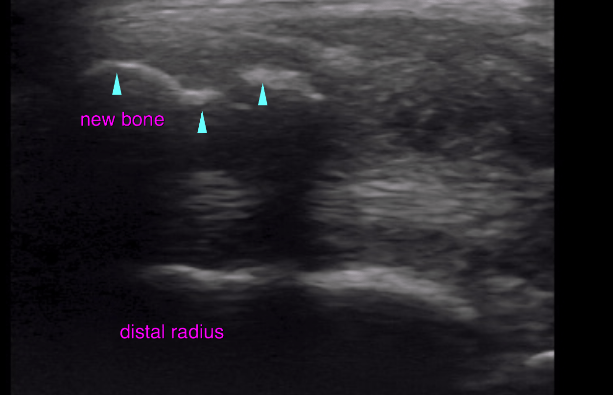
Radiographs: Chronic periosteal reaction distal radius with concurrent severe focal soft tissue swelling. Poss OSA. FNA of area pending. Mild periarticular remodeling of the caudal aspect of the right shoulder, mineralization associated with the greater tubercle. Joint tap pending
Tnis 6 year old FS Labrador Retriever dog presented with right front lameness
Physical exam: tender right shoulder, swelling right carpus
Radiographs: Chronic periosteal reaction distal radius with concurrent severe focal soft tissue swelling. Poss OSA. FNA of area pending. Mild periarticular remodeling of the caudal aspect of the right shoulder, mineralization associated with the greater tubercle. Joint tap pending