A 9-year-old FS mixed breed dog presented for evaluation of episodic coughing that appeared to be somewhat antibiotic responsive. Survey radiographs showed a normal cardiac silhouette, mild interstitial lung pattern, and an irregular shadow superimposing on the craniobasal cardiac silhouette and adherent to the left thoracic wall from the second to the forth rib. The radiographic changes had been present for almost 2 years.
A 9-year-old FS mixed breed dog presented for evaluation of episodic coughing that appeared to be somewhat antibiotic responsive. Survey radiographs showed a normal cardiac silhouette, mild interstitial lung pattern, and an irregular shadow superimposing on the craniobasal cardiac silhouette and adherent to the left thoracic wall from the second to the forth rib. The radiographic changes had been present for almost 2 years.
Case Study
Mild pulmonary hypertension in a 9 year old FS mixed breed dog
Sonographic Differential Diagnosis
Normal heart (in terms of structure and function), but there may be mild pulmonary hypertension. The structure seen on radiography and ultrasound may be fat or fibrous tissue. Neoplasia is less likely but possible.
Image Interpretation
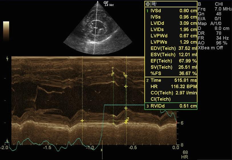
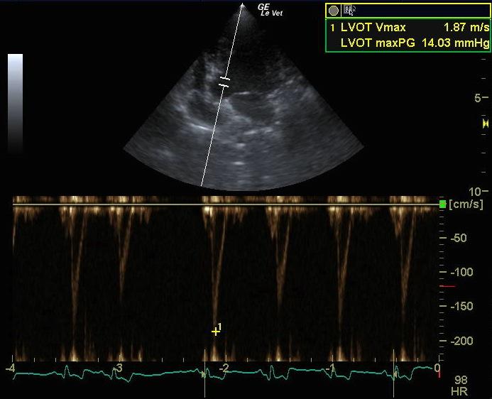
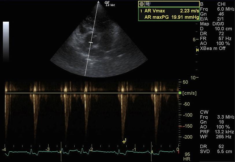
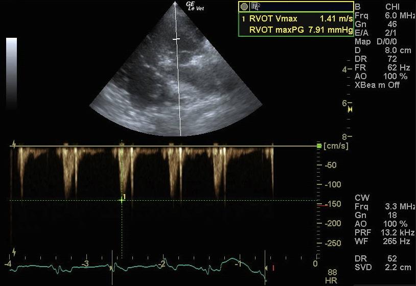
There is a slightly thickened mitral valve, which seems to be properly coadapting during systole. The right ventricle is slightly concentrically hypertrophied, and the right atrium is normal. The septal leaflet of the tricuspid valve is mildly degenerated but no insufficiency is apparent. Flow across the left ventricular outflow tract (LVOT) is mildly accelerated as seen on the continuous wave (CW) profile (2.23 m/s). Flow across the right ventricular outflow tract (RVOT) is normal except for some asymmetry of the flow profile which could, in combination with the slight concentric hypertrophy seen on 2D, be a sign of mild pulmonary hypertension. Ultrasound examination of the structure seen on the radiographs shows some hypoechoic homogenous tissue which is most likely either fat or an organized hematoma/fibrous tissue.
DX
Outcome
Ultrasound-guided fine needle aspirate would be low risk, and recommended, in order to determine the etiology of the thoracic mass. However, it is unlikely that the complaints of the patient (coughing) are related to that structure. A bronchoscopy would be indicated, and sampling for pathohistologic examination would be recommended (cyto brush, lavage).
Clinical Differential Diagnosis
Cardiac – mitral/tricuspid endocardiosis, vegetative endocarditis, myocardial weakness, pericardial effusion, neoplasia. Tracheo-bronchial – bronchiectasis, tracheal collapse, chronic bronchitis, allergic tracheitis, chronic bacterial infection, environmental (dust, tobacco smoke, chemicals), obesity, neoplasia. Pulmonary – neoplasia, fibrosis, pulmonary hypertension. Thoracic mass – neoplasia, lipoma, lymph node, hematoma, granuloma.
Sampling
None
Video
Patient Information
Clinical Signs
- Atelectasis
- Coughing
Images
Clinical Signs
- Atelectasis
- Coughing