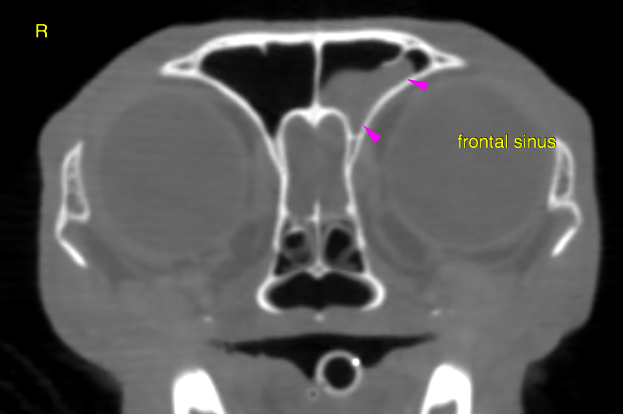
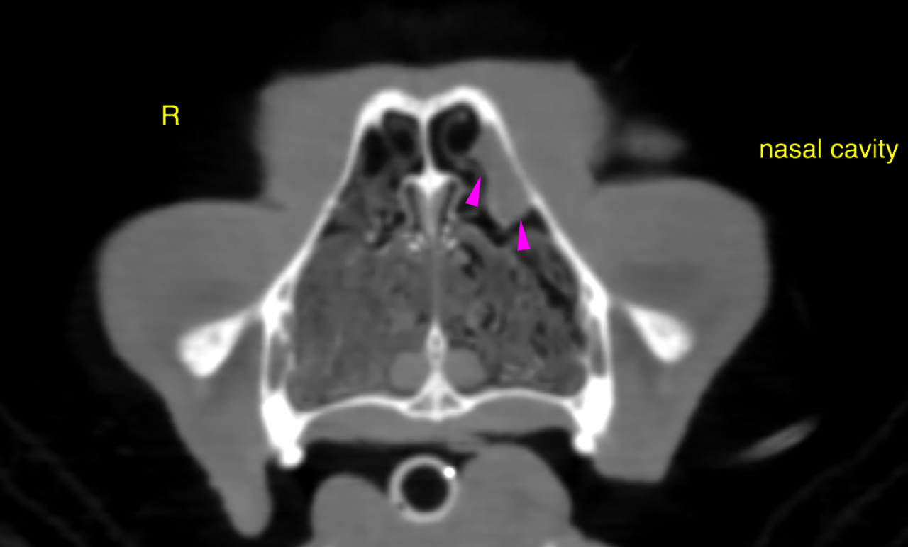
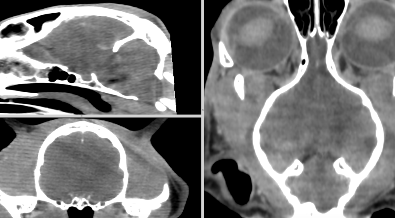
This 9 year old M DSH cat presented with acute onset ataxia and falling to the left. Pysical exam was otherwise wnl. Some improvement with prednisone
This 9 year old M DSH cat presented with acute onset ataxia and falling to the left. Pysical exam was otherwise wnl. Some improvement with prednisone