CT – Malignant soft tissue neoplasia with secondary osteolysis of the maxillary, zygomatic, nasal, orbital and palatinal bones in a 8 year old MN Canine

Case Study

CT – Malignant soft tissue neoplasia with secondary osteolysis of the maxillary, zygomatic, nasal, orbital and palatinal bones in a 8 year old MN Canine

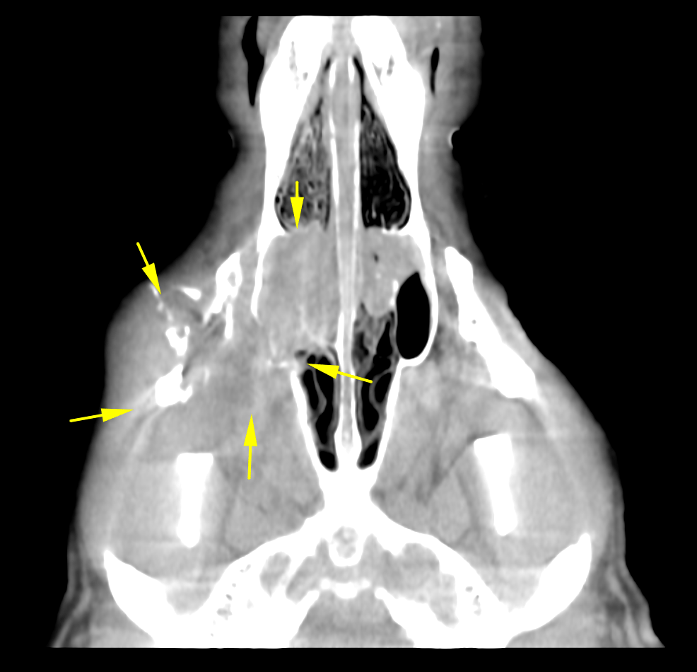
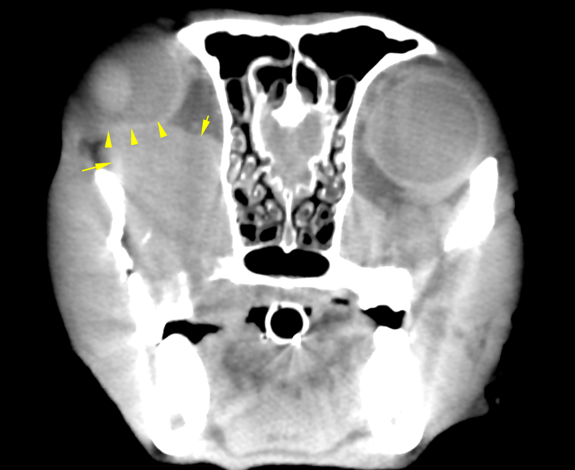
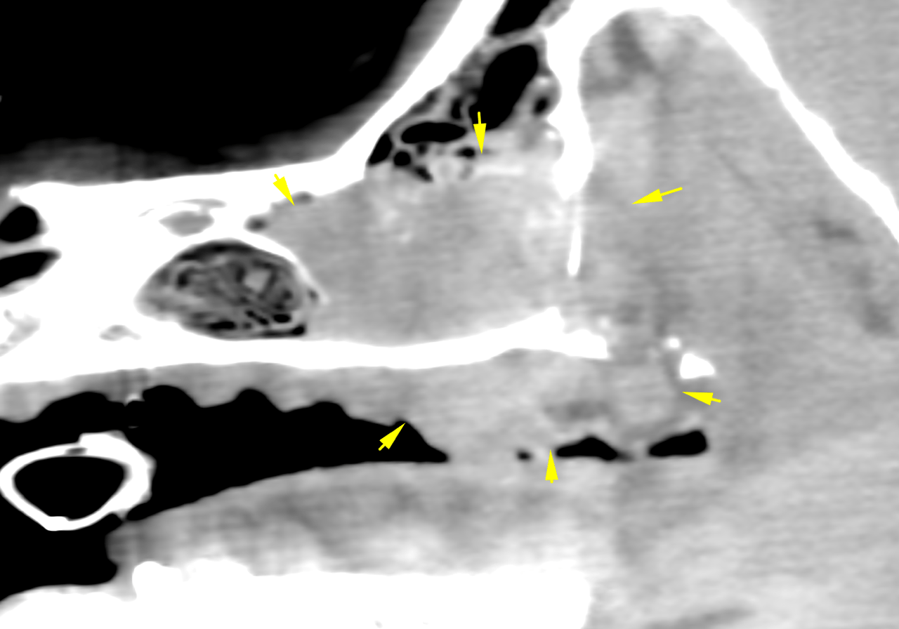
The patient is a 8 year old MN canine presented for a dental cleaning. During the procedure a mass was noted on the right maxilla extending past the dental arcade medially. Right eye bupthalmia. 
The patient is a 8 year old MN canine presented for a dental cleaning. During the procedure a mass was noted on the right maxilla extending past the dental arcade medially. Right eye bupthalmia. 
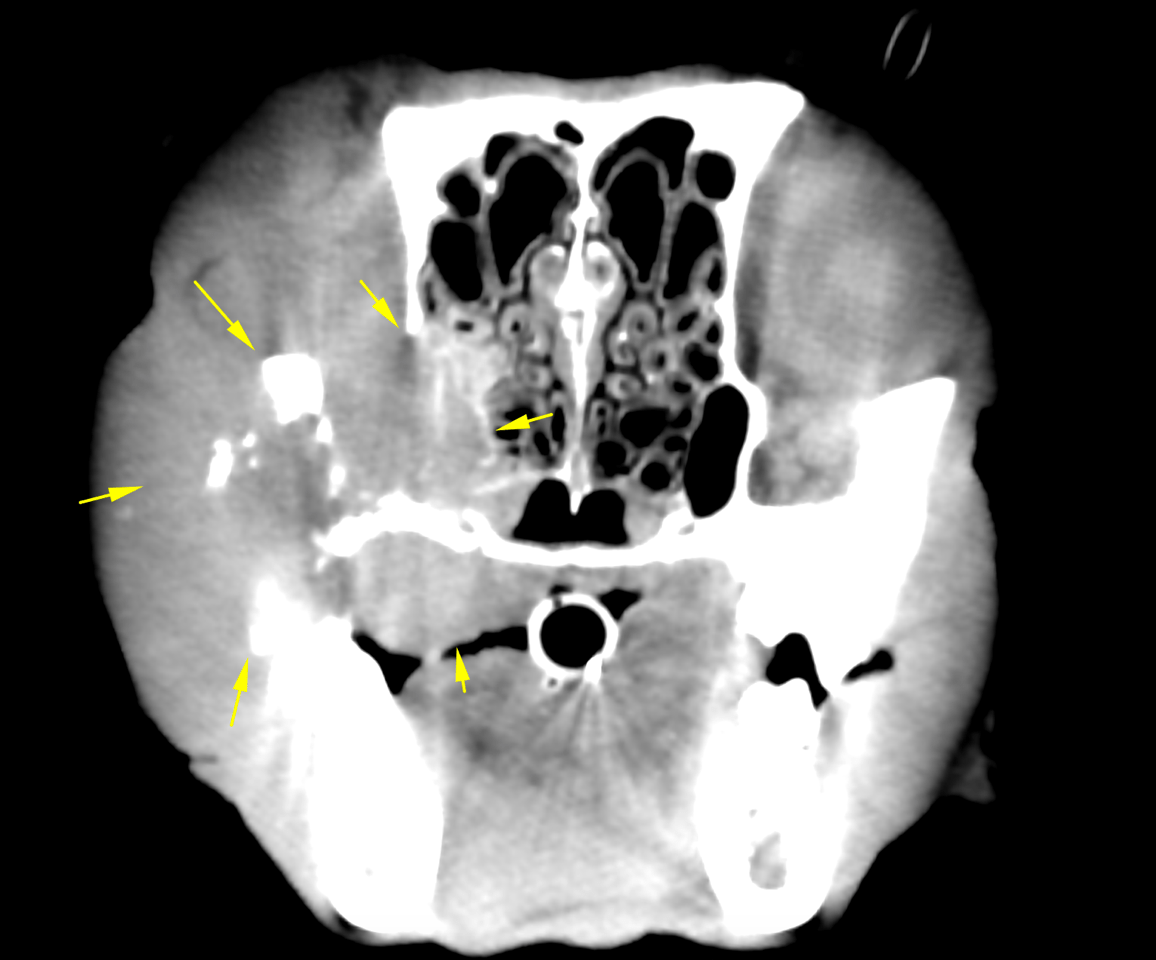
Image Interpretation

CT of the head – A large soft tissue attenuating ovoid mass is seen at the right maxilla along the alveolar
crest caudal to the second premolar (106). Aggressive permeative osteolysis of the
underlying alveolar bone and neighboring maxillary, palatinal and nasal bones as well
as the zygomatic arch and bony orbita is noted. Mild amorphous periosteal reactions of
the bone are seen at the tumoral margins. The mass shows expansile growth and
penetrates into the right nasal and oral cavity as well as into the right orbita causing
turbinate destruction and exophthalmus with a mass effect on the bulbus. The teeth 107
to 110 reveal severe displacement owing to the mass effect of the lesion and loss of
their alveolar bone.
Increase of nasal secretions and mucosal thickening of the nasal turbinates is noted
within the right nasal cavity.
The submandibular lymph nodes are within normal limits. The medial retropharyngeal
lymph nodes are not included in the scan.
There are bilateral signs of healed chronic otitis.

DX

Malignant soft tissue neoplasia of the maxillary, zygomatic, nasal, orbital and palatial bones with turbinate destruction

Outcome

Differential diagnoses for the mass include squamous cell carcinoma, melanoma,
fibrosarcoma, and lymphosarcoma as well as acanthomatous epuli.
Tumor resection would require complete leftsided hemimaxillectomy, resection of the
bony orbita and orbital eventration. The prognosis for curative surgery is poor.
Palliative cytoreducing surgery with adjuvant radiation or chemotherapy may be
considered.
Complete staging with aspiration of the regional lymph nodes to rule out metastatic
spread 3 view chest radiographs and ultrasound of the abdomen.

Patient Information

Patient Name : Gizmo Richards
Gender : Male, Neutered
Species : Canine
Type of Imaging : Ultrasound
Status : Complete
Liz Wuz Here : Yes

Exam Finding

  • Masses
  • Ocular abnormality

Images

bildschirmfoto_2015-12-23_um_12bildschirmfoto_2015-12-23_um_12bildschirmfoto_2015-12-23_um_12bildschirmfoto_2015-12-23_um_12