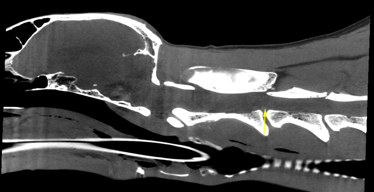
The patient is a 8 year old FS Greyhound dog presented with tetra paresis. Started out with right front leg held stiffly on 2 days ago. Yesterday she held her head down and it seemed to bob a bit. Not nearly as active. Taken to rDVM—BW was done and per O, diagnosed with neck injury (painful when head turned to the left). Given Deramaxx and sent home with Robaxin. Today could not stand.
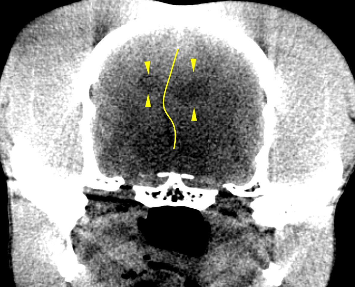
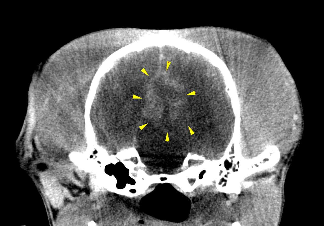
CP deficits to all 4 paws (none seen). Will try to support weight with all paws except front right. Superficial pain present, anal tone present. CARDIOVASCULAR-1/6 left sided murmur
The patient is a 8 year old FS Greyhound dog presented with tetra paresis. Started out with right front leg held stiffly on 2 days ago. Yesterday she held her head down and it seemed to bob a bit. Not nearly as active. Taken to rDVM—BW was done and per O, diagnosed with neck injury (painful when head turned to the left). Given Deramaxx and sent home with Robaxin. Today could not stand.
CP deficits to all 4 paws (none seen). Will try to support weight with all paws except front right. Superficial pain present, anal tone present. CARDIOVASCULAR-1/6 left sided murmur