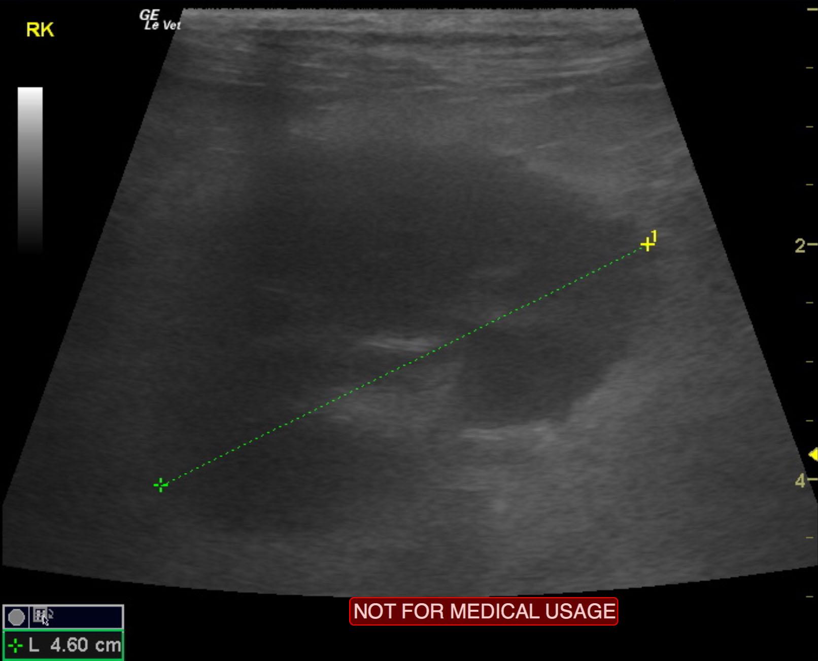
A 13-year-old spayed female DLH cat was presented for evaluation of recent onset poorly controlled diabetes mellitus. Abnormalities on laboratory workup included urinary tract infection, leukocytosis with a left shift, hyperglycemia, elevated BUN and elevated phosphorus. CPL was negative.
A 13-year-old spayed female DLH cat was presented for evaluation of recent onset poorly controlled diabetes mellitus. Abnormalities on laboratory workup included urinary tract infection, leukocytosis with a left shift, hyperglycemia, elevated BUN and elevated phosphorus. CPL was negative.