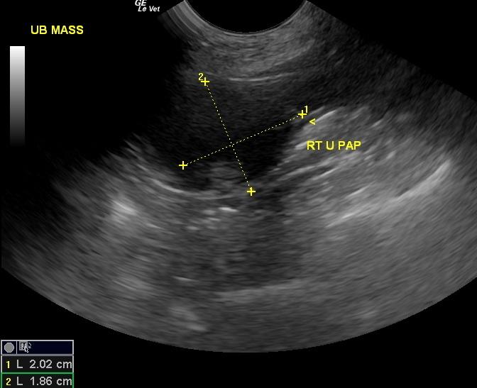
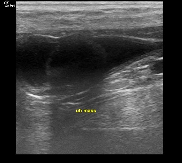
A 10-year-old spayed female Boston Terrier was presented for intermittent hematuria. Physical examination was unremarkable. The patient was treated with a course of antibiotics, pending urinalysis and urine culture. On urinalysis, the urine was cloudy and amber in appearance, the specific gravity was normal, and there was 1+ bilirubinuria, 2+ hematuria, leukocyturia, and mild triple phosphate crystalluria. Urine culture was negative.
A 10-year-old spayed female Boston Terrier was presented for intermittent hematuria. Physical examination was unremarkable. The patient was treated with a course of antibiotics, pending urinalysis and urine culture. On urinalysis, the urine was cloudy and amber in appearance, the specific gravity was normal, and there was 1+ bilirubinuria, 2+ hematuria, leukocyturia, and mild triple phosphate crystalluria. Urine culture was negative. On recheck urinalysis two weeks later, the urine was clear and light yellow in appearance, the specific gravity was normal and there was 1+ proteinuria and 3+ hematuria. Survey radiographs initially showed a possible urethral calculus above the descending colon. However, a radiographic consult revealed only degenerative spinal changes, no definitive evidence of urinary tract calculi, and a focal 4mm diameter ovoid soft-tissue opacity between the descending colon and the ventral aspect of L6.