An 11-year-old FS DSH with history of asthma was presented for acute hind leg paresis. Abnormalities on physical examination were tachycardic, pronounced gallop rhythm, absent femoral pulses, all four extremities were cool, and she was unable to stand. The patient was treated with Lasix, nitroglycerin, and heparin. Within a few hours she was able to ambulate on her own but was knuckling over on all fours
An 11-year-old FS DSH with history of asthma was presented for acute hind leg paresis. Abnormalities on physical examination were tachycardic, pronounced gallop rhythm, absent femoral pulses, all four extremities were cool, and she was unable to stand. The patient was treated with Lasix, nitroglycerin, and heparin. Within a few hours she was able to ambulate on her own but was knuckling over on all fours
Case Study
15-00022 Hannah C Cardiomyopathy ———NO IMAGES—–
Sonographic Differential Diagnosis
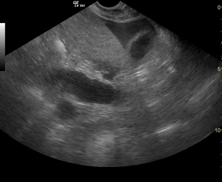
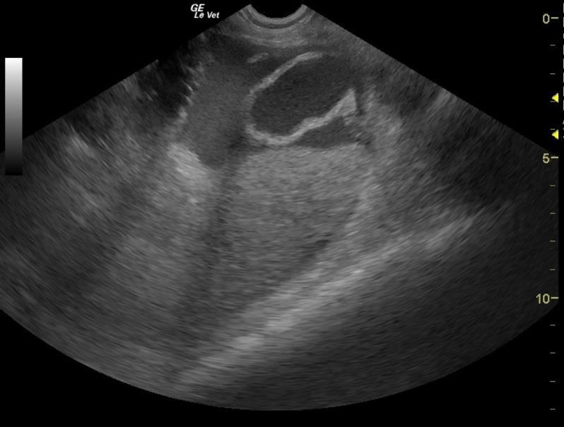
Cardiomyopathy which is either consistent with mild HCM with systolic dysfunction or restrictive cardiomyopathy / intermediate cardiomyopathy. The enlarged left atrium is certainly consistent with the possibility of an embolism originating from primary cardiac disease although there are no visible thrombi in the atria (this certainly does not rule out thromboembolic disease).
Image Interpretation
The echocardiogram revealed hypertrophic cardiomyopathy. The left ventricular free wall and septal wall were both measured at the high end of normal to mildly thickened at 0.58cm each. The papillary muscles were prominent. The fractional shortening was decreased indicating there is some degree of systolic dysfunction. Mitral regurgitation was present. There was no systolic anterior motion of the mitral valve and there was no obvious thrombi. The left atrium was moderately to severely enlarged in size. There was no evidence of any thrombi or smoke in the left atrium. The right side of the heart is normal in appearance. Pulmonic flows and aortic flows were within normal limits. There was no evidence of pleural or pericardial effusion nor any masses.
DX
Outcome
The patient was recommended for thoracic radiographs, blood work prior to treatment with cardiac medications, EKG, and a blood pressure measurement. Before any assessment or therapy was done she died on her own overnight. Therapy would have included Enalapril, Plavix, low dose aspirin, and heparin; as well as theophylline and an inhalant steroid for the asthma.
Clinical Differential Diagnosis
Aortic thrombo-embolism secondary to cardiac disease, toxicosis, trauma, shock
Sampling
None taken.
Video
Patient Information
Clinical Signs
- Weakness
Exam Finding
- Cool foot
- Gallop rhythm
- Pulse deficits
- Tachycardia
- Weakness
Images
Clinical Signs
- Weakness