08_00157 Harley R Splenic hematoma, puppy HBC

Case Study

08_00157 Harley R Splenic hematoma, puppy HBC

A 7-month-old SF Pit bull terrier was presented for evaluation of dysuria and intermittent hematuria following a car accident.  Urinalysis showed an SG of 1.012 and hematuria. CBC and serum biochemistry were within reference range. Survey radiographs showed a normal thorax and loss of abdominal detail. 
 

Sonographic Differential Diagnosis

Swollen kidneys, renal cyst.
Ruptured spleen with hematoma and hemoabdomen.

Image Interpretation

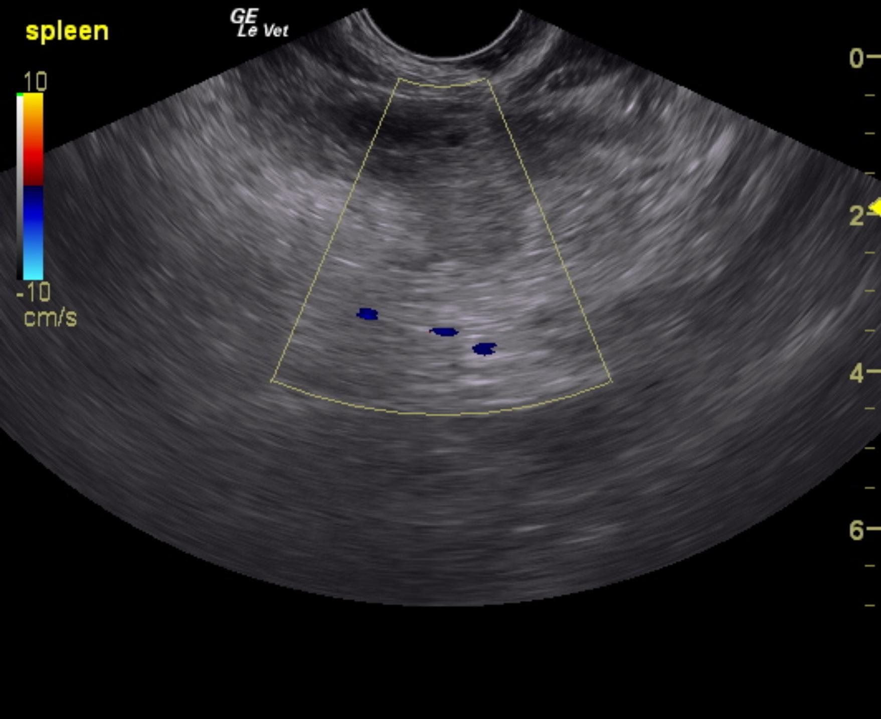
The spleen presented a hematoma that measured approximately 3.0 cm at the mid caudal body with ill-defined surrounding omentum. This appears to be stable.
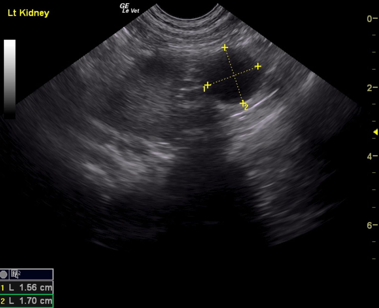
Anechoic cysts were noted in the caudal pole of the left kidney measuring 1.7 x 1.56 cm. The remainder of the left kidney was unremarkable.
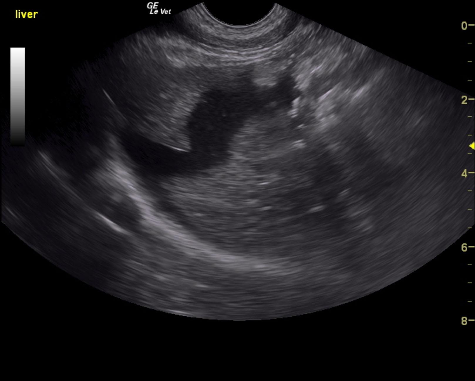
Free fluid was noted in the abdomen around the liver and spleen. Reactive mesentery was noted throughout the abdomen.

DX

Ruptured spleen with hematoma and hemoabdomen.

Outcome

None

Clinical Differential Diagnosis

Intra-abdominal hemorrhage – spleen, liver, kidney, mesentery
Bladder rupture

Sampling

None

UA Specific Gravity Range

1.012

Patient Information

Gender : Female, Spayed
Species : Canine
Type of Imaging : Ultrasound
Status : Complete

Clinical Signs

  • Dysuria
  • Hematuria

Images

harl4harl2harl3

Clinical Signs

  • Dysuria
  • Hematuria

Urinalysi

  • Blood Present
  • Specific Gravity Low