08_00156 Einstein M Splenic nodules, presumed mets from ASA

Case Study

08_00156 Einstein M Splenic nodules, presumed mets from ASA

An 8-year-old NM pug with an anal gland adenocarcinoma and possible enlarged sublumbar lymph node was presented for staging.


Sonographic Differential Diagnosis

This is strongly suggestive for metastatic neoplasia likely of anal gland adenocarcinoma origin.

Image Interpretation

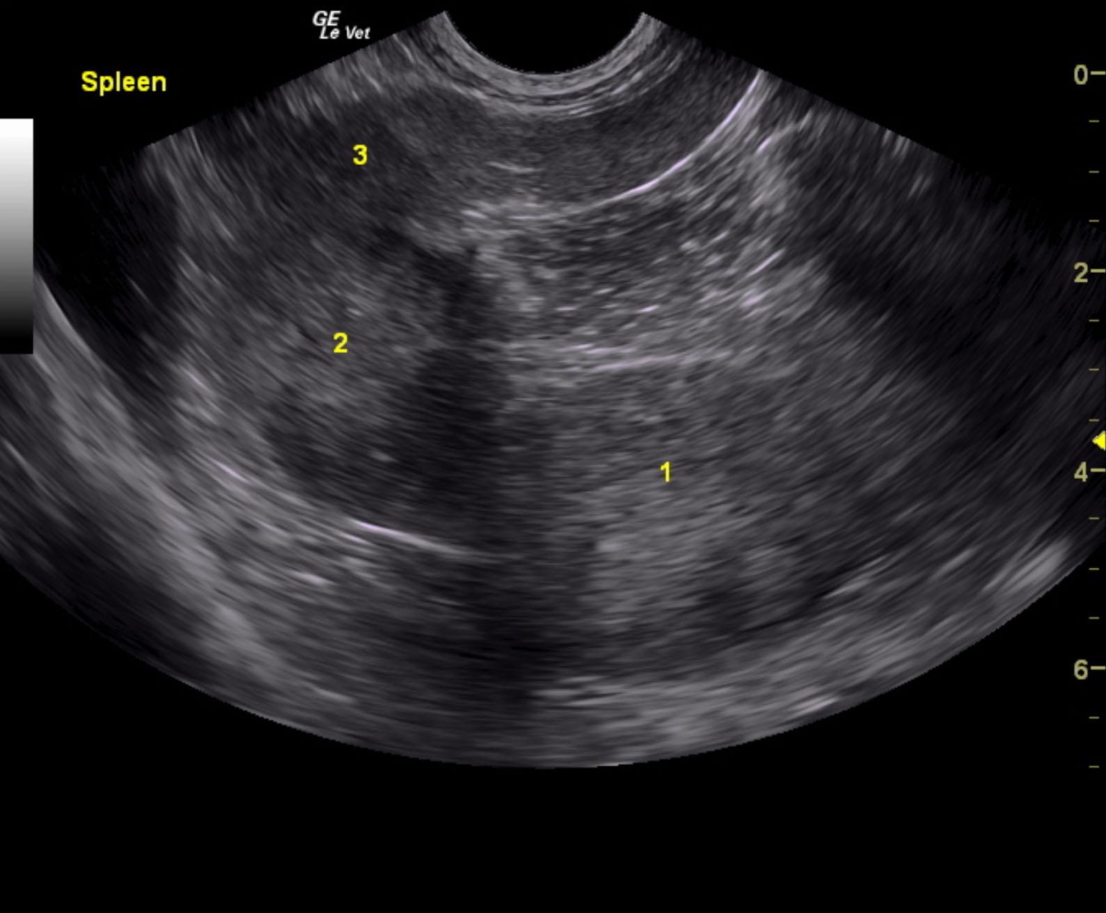
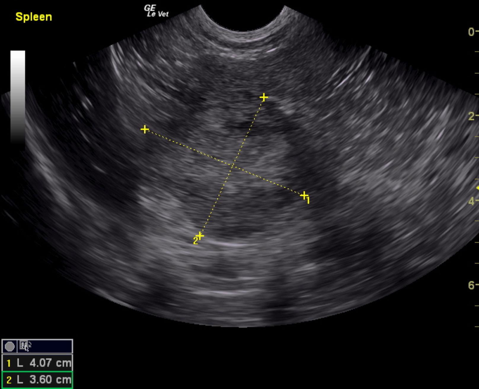
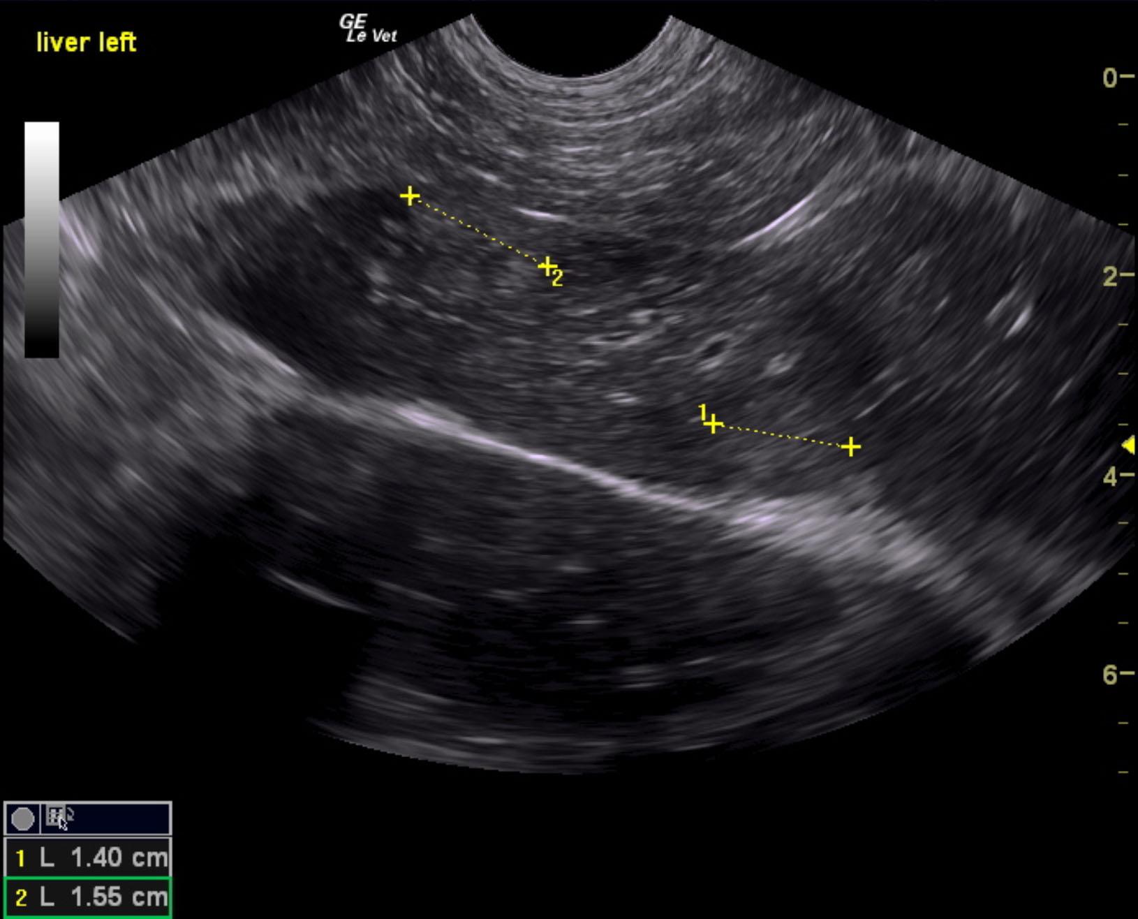
The spleen in this patient presented three separate expansive masses. The masses measured 1.0, 2.0 and 4.07 cm each. These masses were parenchymal enlargements and likely metastatic in nature given the patient’s history. There was no evidence of rupture noted.The liver presented multiple, hyperechoic nodules with some disruption of architecture measuring 1.4 cm and 1.5 cm in the deep left liver with other heterogenous changes. The internal iliac (hypogastric) lymphnode deviates the descending colon.

DX

Metastatic nodules in spleen

Outcome

None

Clinical Differential Diagnosis

Metastatic neoplasia – sub-lumbar lymph nodes, spleen, liver

Sampling

None

Patient Information

Gender : Male, Neutered
Species : Feline
Type of Imaging : Ultrasound
Status : Complete

Exam Finding

  • Lymphadenopathy

Images

einst1ein2ein4
Skip to content