A 12-year-old SF Scottish terrier was presented for evaluation of chronic UTI. Urinalysis showed a specific gravity of 1.013 and hematuria.
A 12-year-old SF Scottish terrier was presented for evaluation of chronic UTI. Urinalysis showed a specific gravity of 1.013 and hematuria.
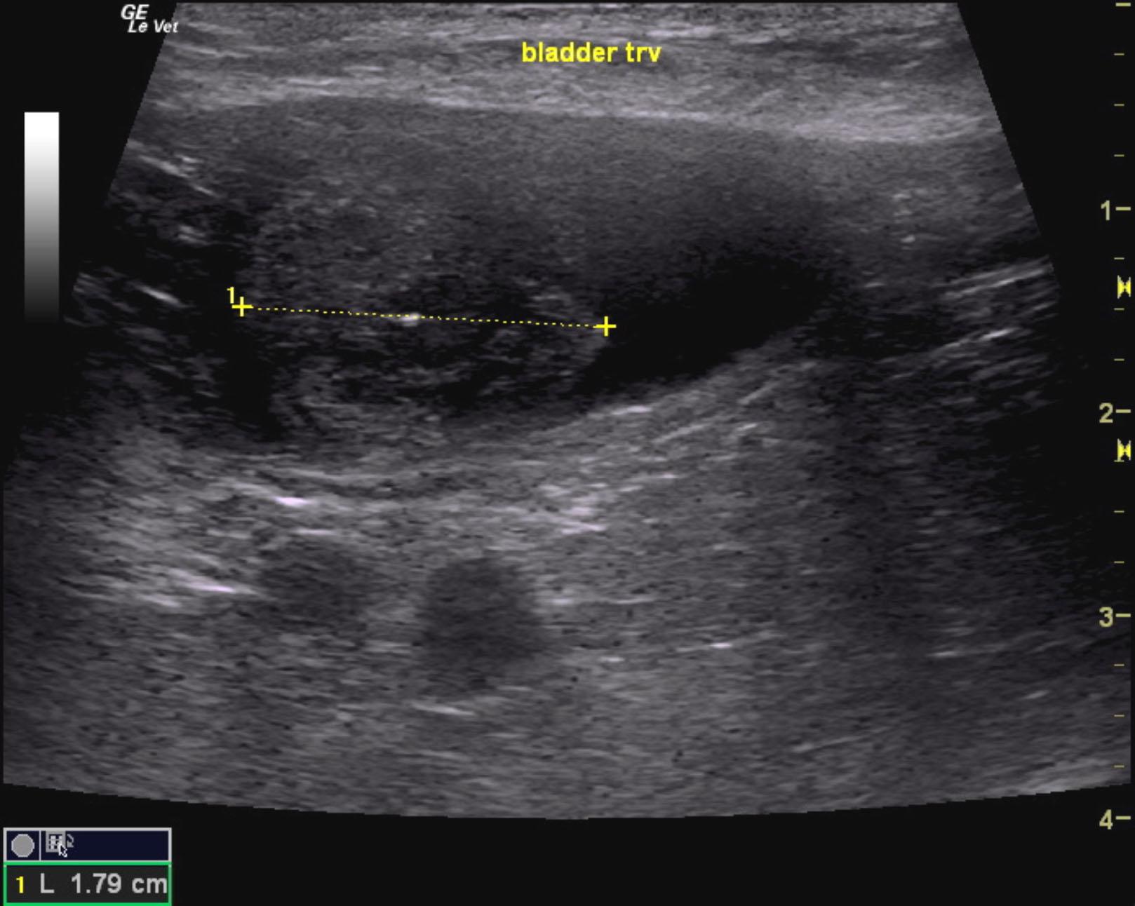
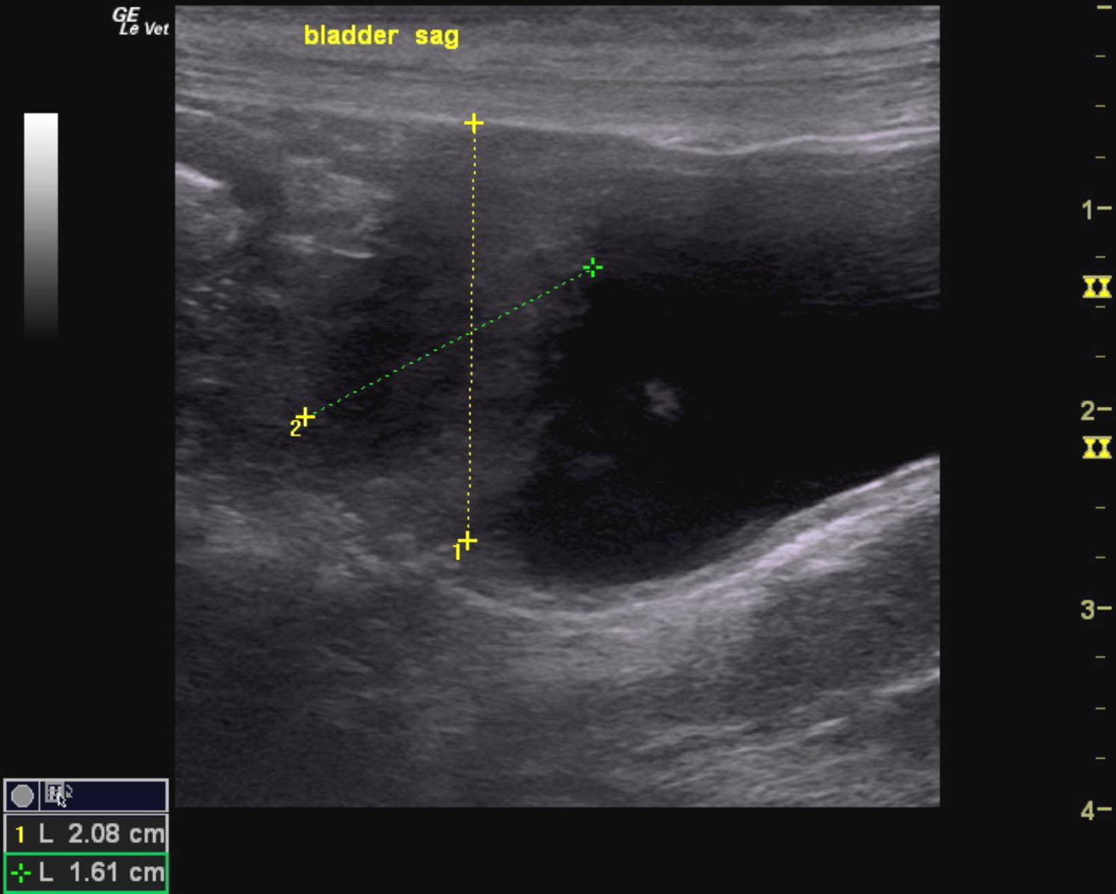
Bladder mass.
Recommend surgical resection in this patient removing the cranial half of the urinary bladder to ensure that adequate borders are achieved. Otherwise, intraoperative ultrasound to delineate the normal wall can also be considered. There is a minor potential for chronic cystitis in this patient instead of transitional cell carcinoma. However, the pattern is most consistent with TCC and resection is essential.
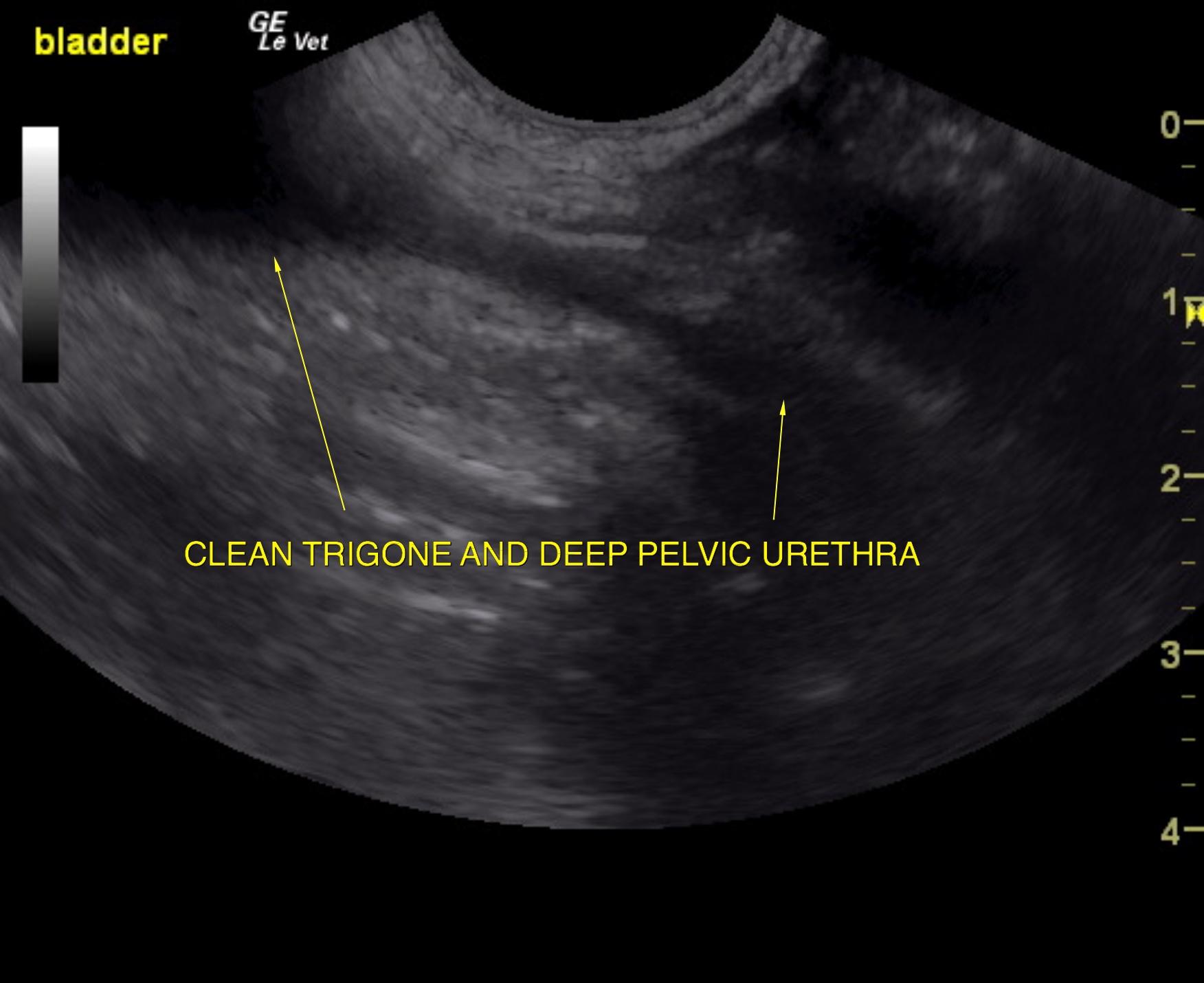
The urinary bladder presented a 1.8 cm mineralizing mass. The bladder mass was apical and appears resectable. The pelvic urethra was imaged 3.0 cm beyond the cystourethral junction and was structurally unremarkable as was the trigone. No obstructive pattern was noted. The apex of the bladder wall was also thickened with loss of detail and measured 2.08 x 1.6 cm.
None
Bladder – chronic bacterial cystitis, uroliths, neoplasia, polyploid cystitis
Kidney – neoplasia, renolith, pyelonephritis
None