A 10-year-old NM Golden retriever was presented for evaluation of hematuria. The only abnormality on urinalysis was the presence of RBC’s.
A 10-year-old NM Golden retriever was presented for evaluation of hematuria. The only abnormality on urinalysis was the presence of RBC’s.
Hydronephrosis and left ureteral mass, potentially of adrenal gland origin. Secondary hydronephrosis. Minor degenerative changes in the right kidney. Echogenic debris in the bladder with cystitis pattern.
The urinary bladder presented echogenic debris and micropolypoid changes. This is consistent with chronic cystitis. The pelvic urethra was unremarkable. The distal ureters were not visible, which is normal.
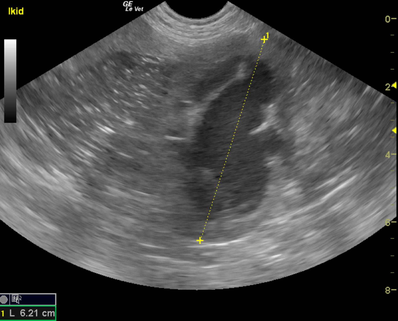
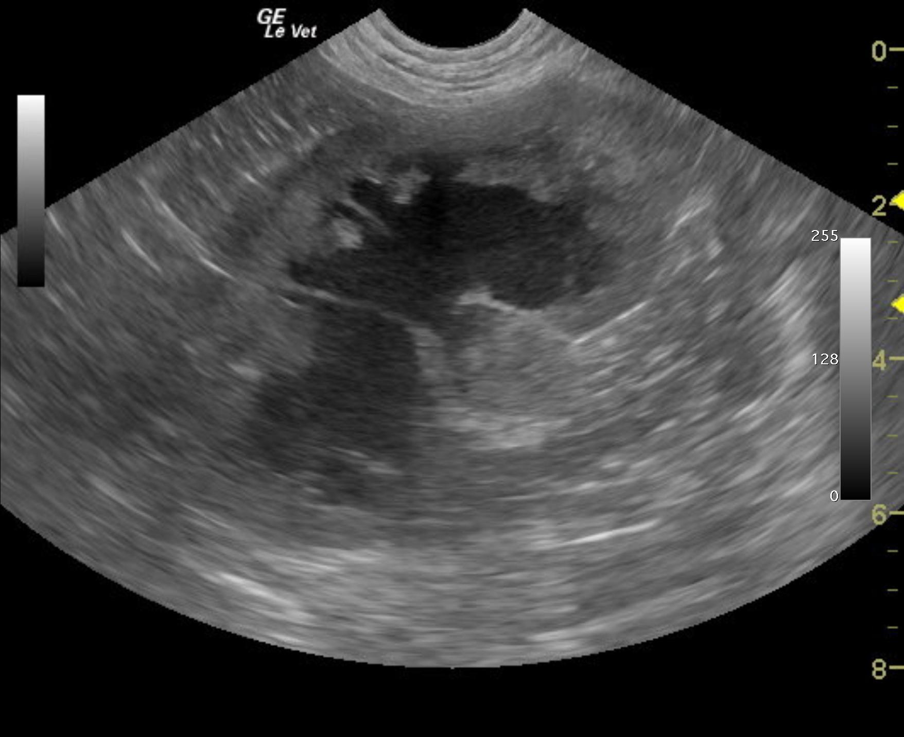
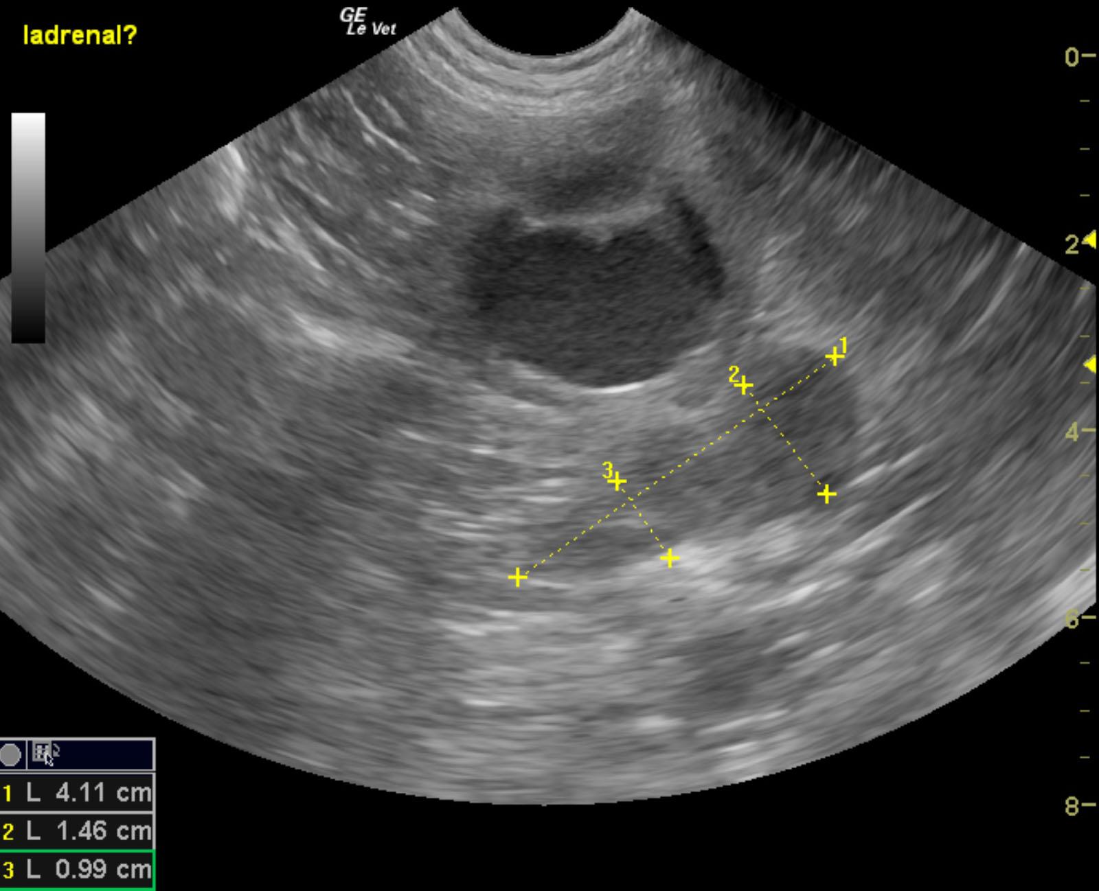
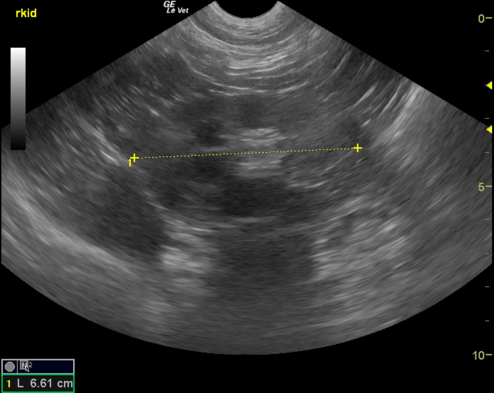
The left kidney presented moderate hydronephrosis with echogenic debris and a pericapsular inflammatory pattern. The left kidney was subnormal in size and measured 6.21 cm. A mass, which appeared to be deriving from the left adrenal gland appears to be causing a stricture of the ureter or the mass could be deriving from the ureter itself. This caused hydronephrosis and outflow obstruction. The right kidney was also subnormal in size and measured 6.61 cm with minor degenerative changes.
None
Bladder – uroliths, neoplasia, granulomatous cystitis, polyploid cystitis, bacterial cystitis
Prostate – neoplasia, prostatitis
Renal – renolith, pyelonephritis, neoplasia
None