06_00231 Clarence C Interstitial cystitis

Case Study

06_00231 Clarence C Interstitial cystitis

A 3-year-old NM DSH was presented for evaluation of hematuria with a history of a previous episode with stranguria a month ago. 

A 3-year-old NM DSH was presented for evaluation of hematuria with a history of a previous episode with stranguria a month ago. 

Sonographic Differential Diagnosis

Interstitial cystitis. Potential concurrent urinary tract infection. Full thickness bladder wall biopsy is recommended.
Interstitial nephrosis with pyelectasia. Renal biopsy can also be considered given convenience of the procedure given that the bladder wall biopsy is ideal in this case for long term management. Urine culture and sensitivity, pain measurement, behavioral issues and glucosamine support can all be considered in this patient. I do not recommend cortisone usage unless histopathology of the bladder wall would suggest a lymphoplasmacytic infiltrate. Bladder lymphoma is also a mild potential.
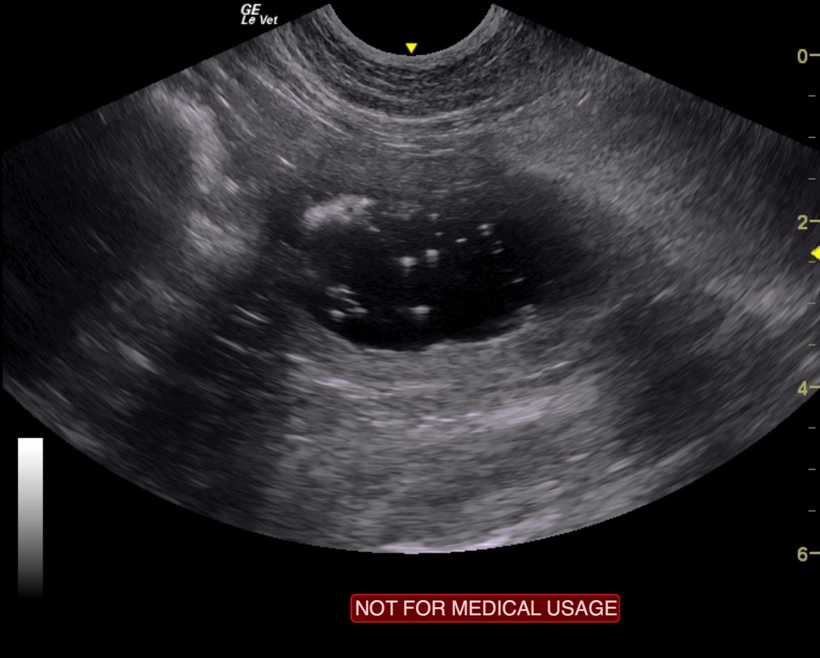
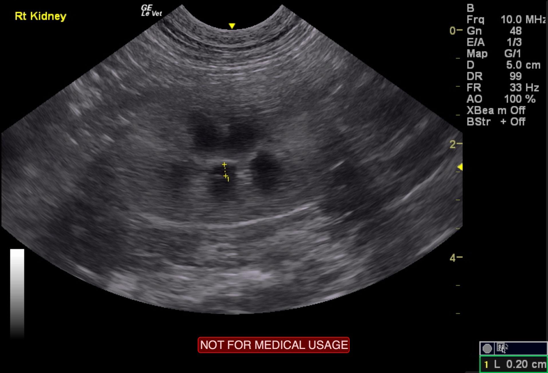
Image Interpretation

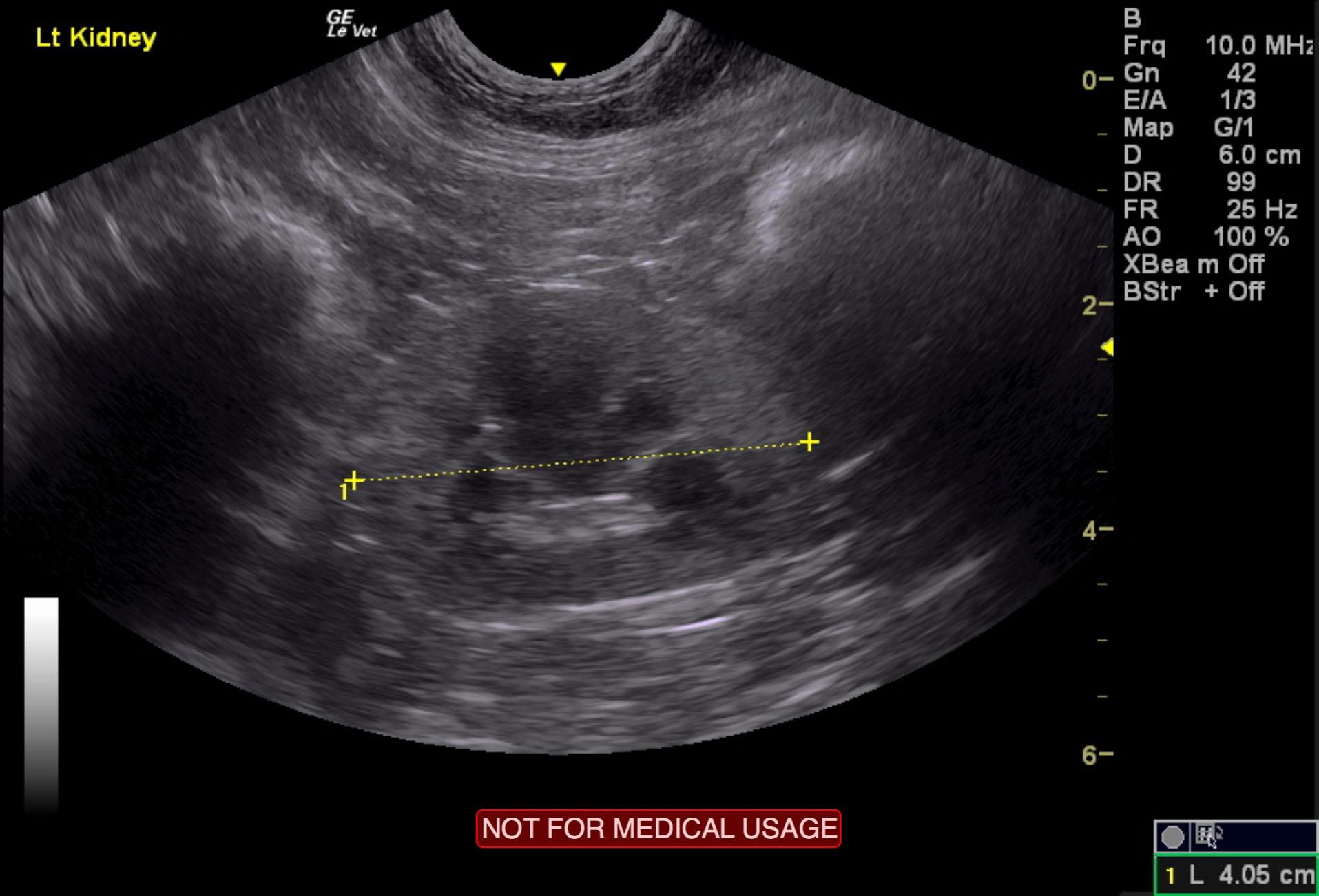
The urinary bladder presented concentric mural thickening with echogenic remodeling of the bladder wall and some loss of detail. Attached mucous was noted adhered to the bladder wall. This is most consistent with interstitial cystitis. There was a minor amount of suspended and dependent sand and debris noted. The bladder wall at moderate repletion measured 0.34 cm. The kidneys presented a relatively uniform cortical hyperechogenicity when compared to the renal medulla, spleen and liver. No overt masses were noted. Some mildly echogenic debris was present in the medulla. Corticomedullary definition was nebulous and the ratio favored the cortex slightly. The ureters were not visible and assumed to be normal. These changes are most consistent with chronic interstitial nephritis yet infiltrative disease could not be entirely ruled out without biopsy though neoplasia is not suspected. There was slight pyelectasia in the right kidney measuring 0.2 cm. Cortical infarcts were noted. The left kidney measured 4.05 cm.

DX

Interstitial cystitis

Outcome

None

Clinical Differential Diagnosis

Interstitial cystitis, bacterial cystitis, uroliths, polyploid cystitis, granulomatous cystitis

Sampling

None

Patient Information

Gender : Male, Neutered
Species : Feline
Type of Imaging : Ultrasound
Status : Complete

Clinical Signs

  • Hematuria

Images

clarence_coarts_3_yr_fel_interstitial_cystitisclarence_coarts_3_yr_fel_interstitial_cystitis_rkclarence_coarts_3_yr_fel_interstitial_cystitis_lk

Clinical Signs

  • Hematuria