A 16-year-old NM Miniature Pinscher was presented for evaluation of hematuria. Abnormalities on serum biochemistry were elevated liver enzyme activity (ALT 222, ALP 1023), cholesterol, and triglycerides.
A 16-year-old NM Miniature Pinscher was presented for evaluation of hematuria. Abnormalities on serum biochemistry were elevated liver enzyme activity (ALT 222, ALP 1023), cholesterol, and triglycerides.
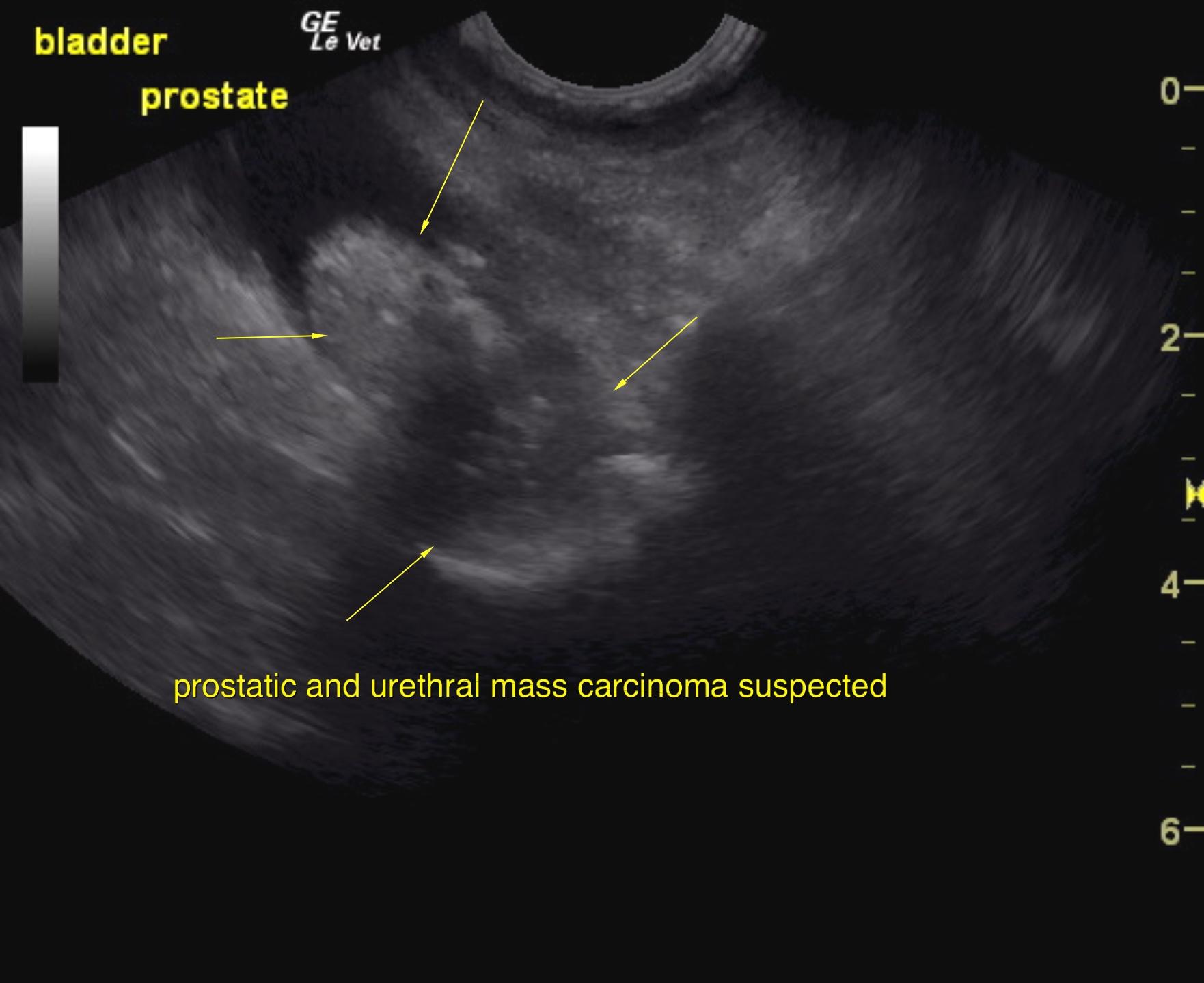
Prostatic and urethral mineralizing mass. Suspect carcinoma.
Concurrent bladder sand.
Traumatic catheterization was performed without complication. Guarded to poor prognosis.
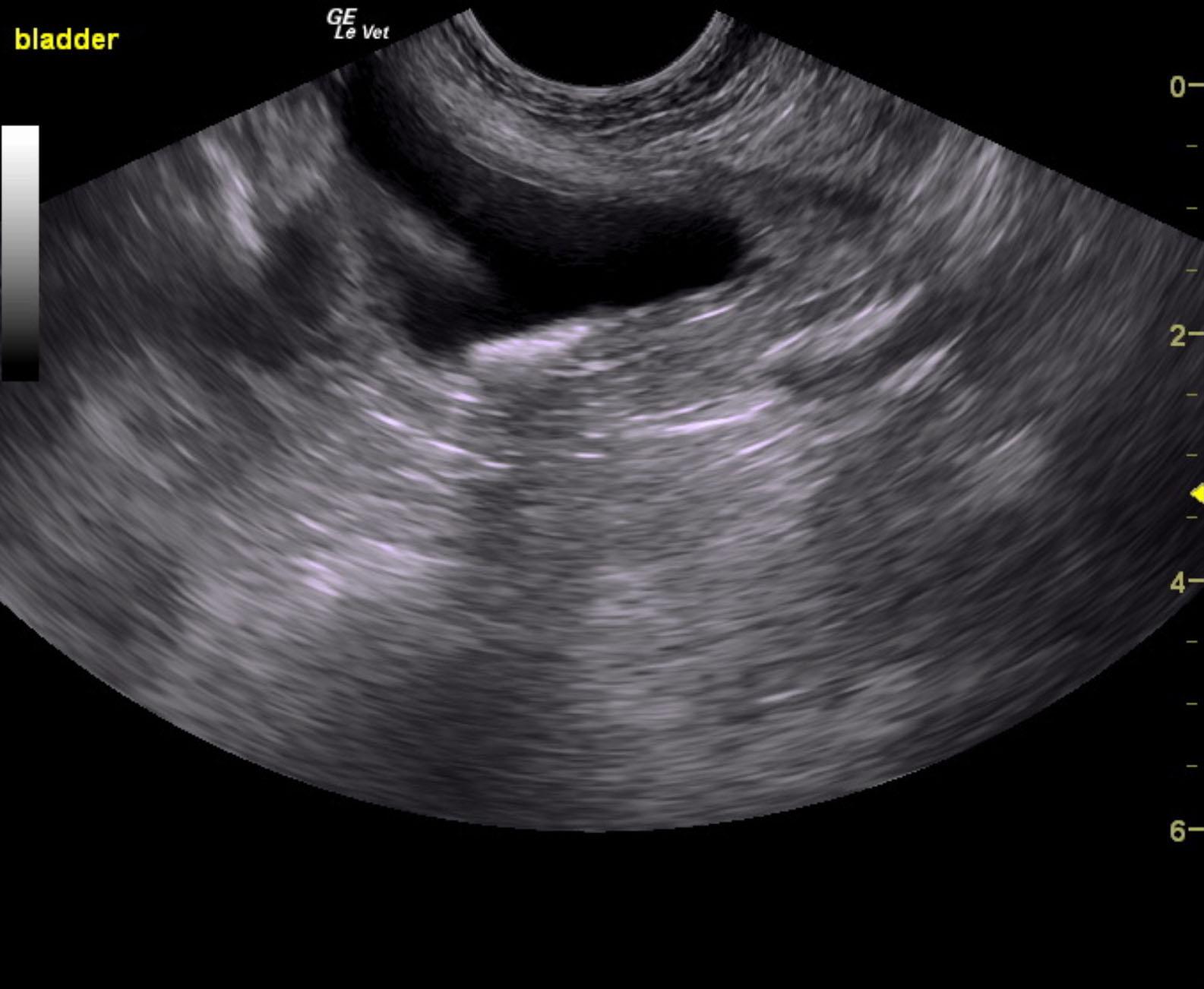
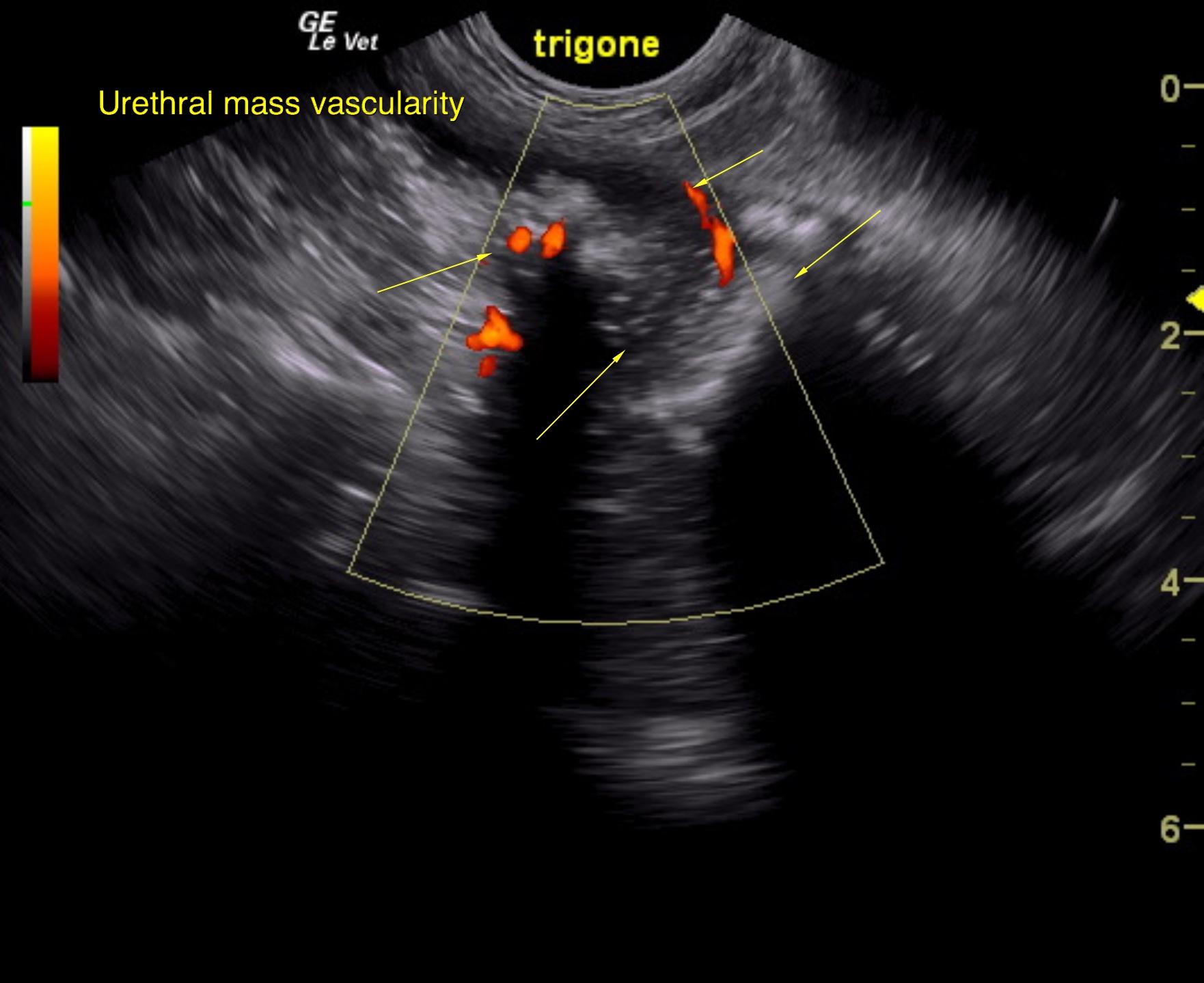
The urinary bladder presented 1.0 cm of sand accumulation. The pelvic urethra was infiltrated with a mineralizing mass. The mass involved the prostate. Power Doppler demonstrates the vascularity of the mass to distinguish it from a collection of debris. Disruption of architecture was noted along with capsular expansion. The infiltrative pattern within the urethra extended past the prostatic urethra.
None
Bladder – neoplasia, uroliths, bacterial cystitis
Prostate – neoplasia, prostatitis
Liver – neoplasia, nodular regeneration, reactive hepatopathy, vacuolar hepatopathy
Gall bladder – mucocele, cholecystitis
Cushing’s disease
??