A 19-year-old NM Corgi with a history GI foreign body surgery was presented for evaluation of vomiting. Survey radiographs and biochemistry were all normal.
Sonographic Differential Diagnosis
Chronic disease, consistent with hypertrophic pyloric gastropathy, is evident in the stomach as well as a concurrent foreign body in the duodenum. There was no evidence of peritonitis noted. However, an immediate exploratory surgery is recommended with expectations towards duodenotomy and gastric biopsy in the level of the pyloric outflow for further definition.
Image Interpretation
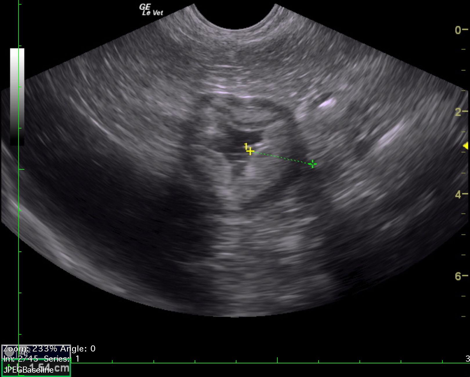
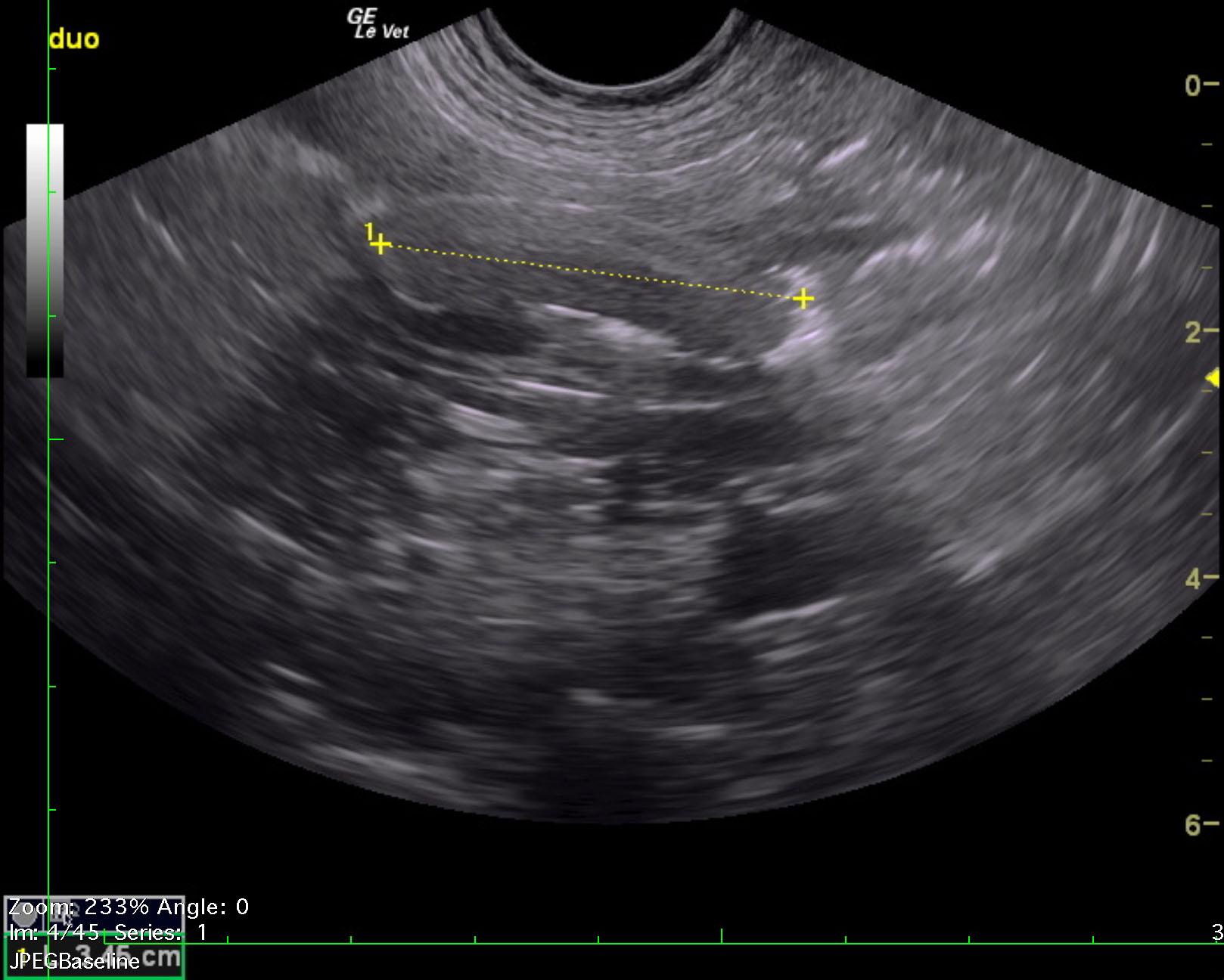
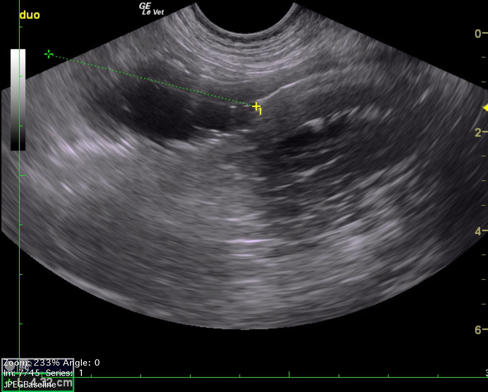
The stomach in this patient presented concentric thickening with hypertrophied echogenic mucosal changes and hypertrophied muscularis. Pyloric wall measured 1.54 cm. The mid duodenum revealed a 3.11 x 0.84 cm isoechoic foreign structure. This is consistent with cork or similar material. An obstructive pattern was noted in the upper duodenum with dilated fluid prior to this foreign structure. The remainder of the intestine was unremarkable.