A 12-year-old NM cat was presented for evaluation of elevated bilirubin (1.11) and ALP (282) and ALT (186) activity.
A 12-year-old NM cat was presented for evaluation of elevated bilirubin (1.11) and ALP (282) and ALT (186) activity.
A 12-year-old NM cat was presented for evaluation of elevated bilirubin (1.11) and ALP (282) and ALT (186) activity.
A 12-year-old NM cat was presented for evaluation of elevated bilirubin (1.11) and ALP (282) and ALT (186) activity.
Suspect hepatic lipidosis with low-grade inflammatory hepatopathy. Possible underlying lymphoma less likely. Primary treatment for lipidosis is recommended in the meantime until cytology results can be assessed.
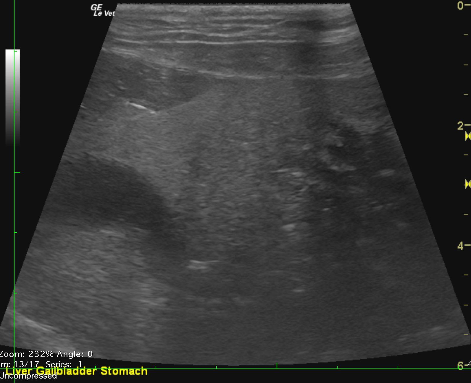
The liver was diffusely hyperechoic compared to the falciform fat. Vascular and biliary tracts were of normal volume and no evidence of congestion was noted. The gallbladder was slightly dilated. This may be owing to anorexia. The cystic duct and common bile duct were slightly tortuous. However, technically the common bile duct was normal in width at the level of the duodenal papilla. The common bile duct measured 0.28 cm. No pathological hepatic lymphadenopathy was evident. No overt structural evidence of inflammatory, infiltrative or regenerative pathology was noted. FNA of the liver were performed without complication.
None
Liver – lipidosis, cholangio-hepatitis, neoplasia, abscessation, granulomatous disease
Gall bladder – obstruction (lith, neoplasia, duodenal/pancreatic disease), cholecystitis
Hemolysis – Mycoplasmosis, IMHA, toxins
Cytology revealed moderate-marked hepatic lipidosis, cholestasis.