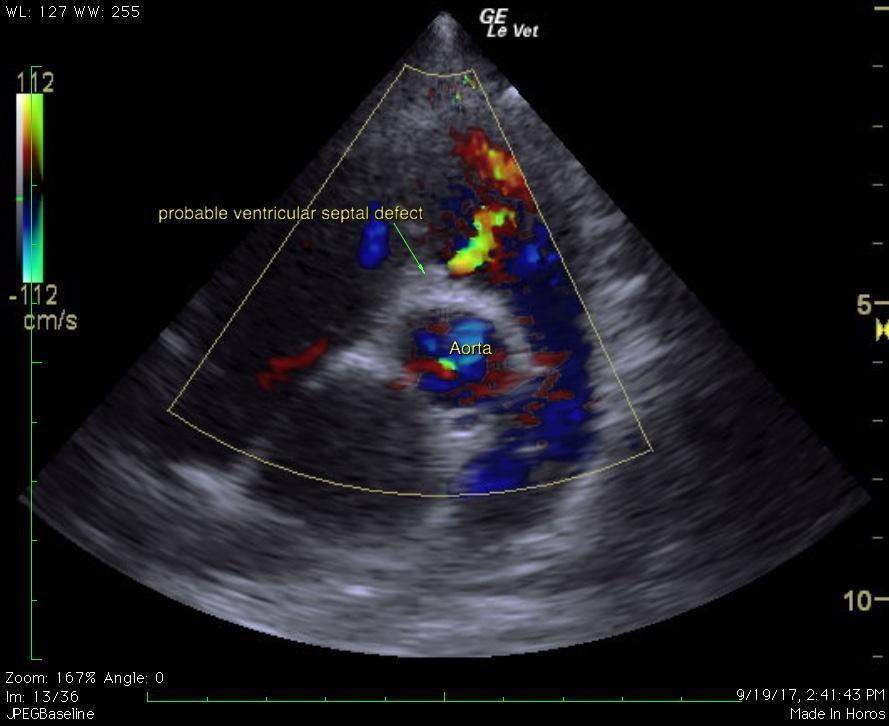
A 1.5-year-old, FS, Boxer was presented for an initial puppy wellness visit and a grade 1/6 cardiac murmur was detected; noted PMI (point of maximal impulse) right cranial. A grade 1-2/6 cardiac murmur was detected at several follow-up visits. The patient underwent ovariohysterectomy without event. More recently the patient was presented for further cardiac evaluations. PE found the patient with a heartrate of 140, panting but with no increased respiratory effort, and synchronous pulses. BP: 111/51, 95/53 MAP 67, 119/66 MAP 78. 2 ECG strips were submitted.
A 1.5-year-old, FS, Boxer was presented for an initial puppy wellness visit and a grade 1/6 cardiac murmur was detected; noted PMI (point of maximal impulse) right cranial. A grade 1-2/6 cardiac murmur was detected at several follow-up visits. The patient underwent ovariohysterectomy without event. More recently the patient was presented for further cardiac evaluations. PE found the patient with a heartrate of 140, panting but with no increased respiratory effort, and synchronous pulses. BP: 111/51, 95/53 MAP 67, 119/66 MAP 78. 2 ECG strips were submitted. The first strip (taken under sedation with butorphanol) showed periods of sinus rhythym and periods that appeared to be a high grade second degree AV block (ventricular rate 40-100 bpm). The second strip showed a sinus rhythm (rate 114 bpm) with intermittent single premature ventricular complexes (RBBB morphology) once sedation had worn off and patient was stimulated.