- 5 year old, 21kg, FS English Bulldog was adopted from a breeding situation in January. She was spayed, received a dental cleaning, nares reduction, and soft palate reduction surgery all under one anesthetic episode. Two months later she presented for a cough and abdominal distension.
- Radiographs read by a radiologist showed a redundant esophagus, aspiration pneumonia, and ascites.
- Bloodwork performed last week shows ALB=2.0g/dL, ALKP=21 U/L, ALT=29 U/L, Chol=347 mg/dL, SDMA=17 mcg/dL, BUN=17mg/dL, Create=1.1mg/dL.
- In house ascitic fluid analysis was consistent with a transudate with occ RBC’s and proteinaceous material. U/A showed proteinuria. The dog was treated with enrofloxacin, Clavamox, and IV fluids.
- The dog presented to this clinic 1 week later for persistent ascites. Echocardiogram shows a normal right heart and a very small MVI. No other insufficiencies are seen. The LA and LA/AO are wnl at 2.51cm and 1.05 respectively.
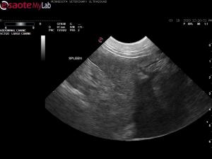
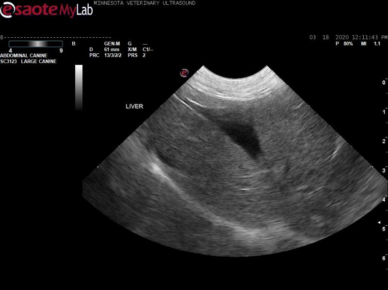
- Abdominal ultrasound shows a moderate amount of free anechoic fluid. There are no visible masses or enlarged lymph nodes. The liver shows increased echogenicity but normal capsule margins, possibly decreased size.
- My differential diagnosis list for the ascites includes chronic liver disease/portal hypertension, infectious disease (mycobacterial, fungal, leptospirosis, rickettsial), vasculitis, thrombus (none seen), and neoplasia. Anything else to put on the list?
- I have recommended a recheck of the ALB, bile acid testing, urinalysis, full ascitic fluid analysis, urine protein:creatinine ratio, Lepto titers, and a 4DX test. If bile acid test results are abnormal, a liver biopsy (surgical, laparoscopic, or ultrasound guided).
- My other differential diagnoses for the decreased ALB include protein losing nephropathy and protein losing enteropathy, however, the ALB is not currently low enough to cause ascites.
- These people may be running out of funds soon. What would you prioritize with this case? Anything else I should be aware of with ascites in a FS English Bulldog?
- Thanks in advance for your input!


Comments
Most likley diagnosis would
Most likley diagnosis would be chronic liver disease with assocaited portal hypertension but need to rule out protein losing nephropathy. Ideal would be a liver biopsy else manage symptomatically.
Update on this case: Bile
Update on this case: Bile acids were normal but UPC came back as 12. Dog is now being managed for PLN. Serum ALB ran at the same time is now 1.6. I have told them to check for Lepto and Lyme but believe owner is out of funds. Still not sure why ascites was present when the ALB was at 2.0. I am thinking either lab error or there was a thrombus in there somewhere.