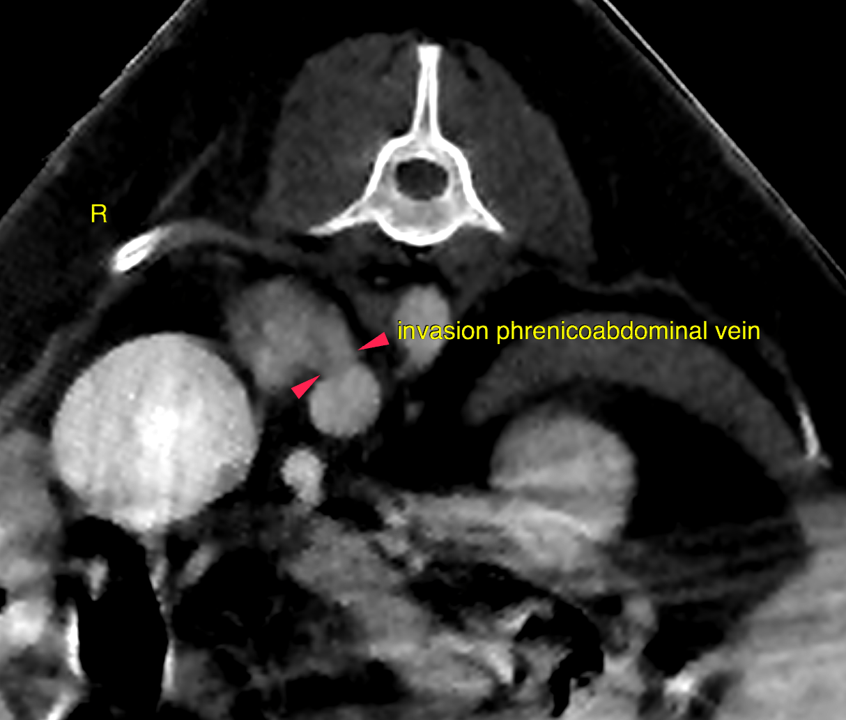
This 12 year old FS Shih Tzu presented for monitoring of a suspected right adrenal mass noted on ultrasound 10 months prior.
CT of the abdomen –
The right adrenal gland shows marked nodular enlargement with rounded shape and
measures 2.5 cm in length, height and width each. There is a tubular extension of 0.5
cm diameter emerging from the craniodorsal aspect of the adrenal mass, which invades
the right phrenicoabdominal vein and has a broad base to the caudal vena cava at their
junction. A mass effect onto the caudal vena cava is noted which leads to focal
leftward displacement of the vessel.
The left adrenal gland is within normal limits for size, shape attenuation and contrast
enhancement.
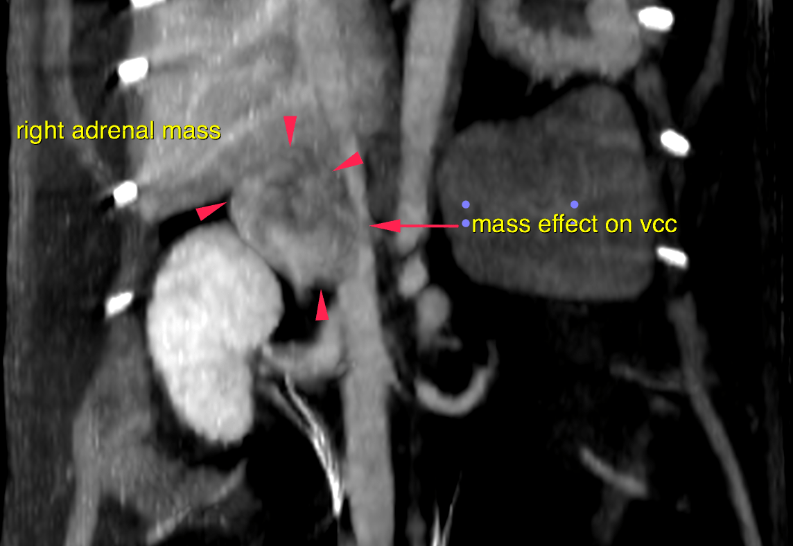
Aggressive
biological behavior of the mass is noted by local vascular invasion into the
phrenicoabdominal vein. The broad contact with and displacement of the caudal vena
cava is consistent with emerging invasion of the caudal vena cava even though a filling
defect is not noted yet. Possible differentials are pheochromocytoma or functional/nonfunctional
adenocarcinoma. Consider complete blood work and urine analysis to check for either
hyperadrenocorticism or metanephrines in the urine in case of pheochromocytoma.
Screening for systemic hypertension is advised as well. Surgical excision of the right
adrenal is advised after completion of the tumor staging