History of a mass on/within the nasal cavity
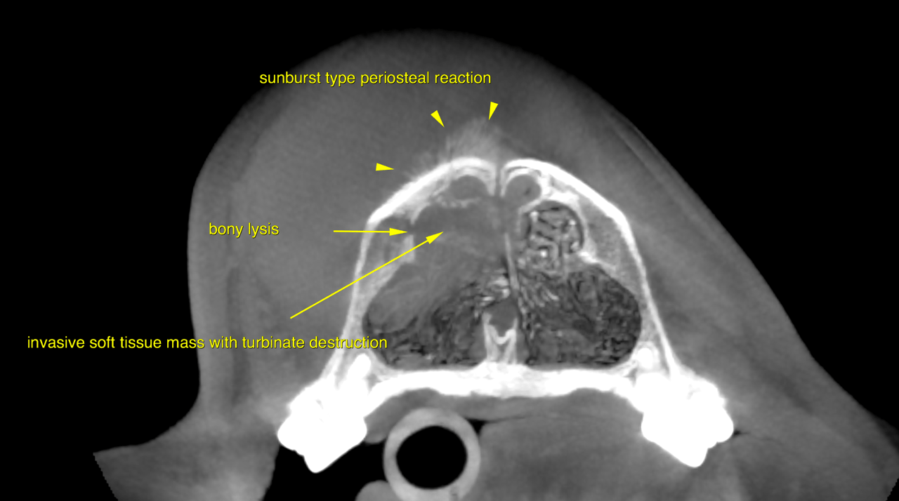
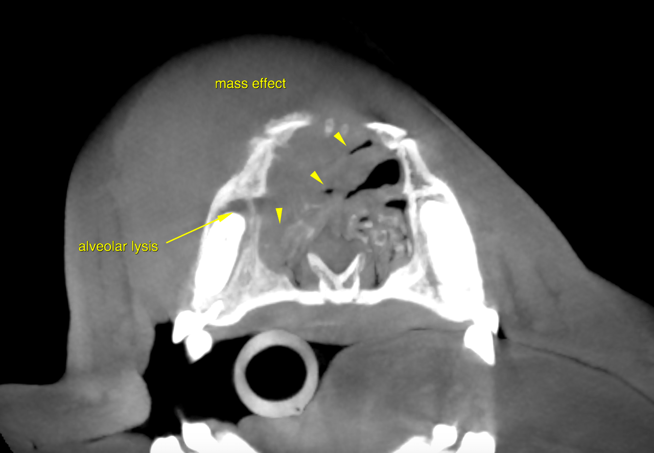
CT of the head, plain and post contrast – A large soft tissue attenuating mass lesion is present on the dorsum of the nose. The
mass measures approximately 5.0 cm in diameter and is positioned to the right of the
midline.
The underlying maxillary and nasal bones reveal multifocal periosteal reactions of
sunburst and amorphous types as well as multifocal permeative osteolysis, which both
are indicators of highly aggressive biological behavior. The soft tissue component of
the mass lesion further presents invasive growth into the right nasal cavity with focal
lysis of the nasal turbinates, as well as lysis of the alveolar bone and dental enamel of
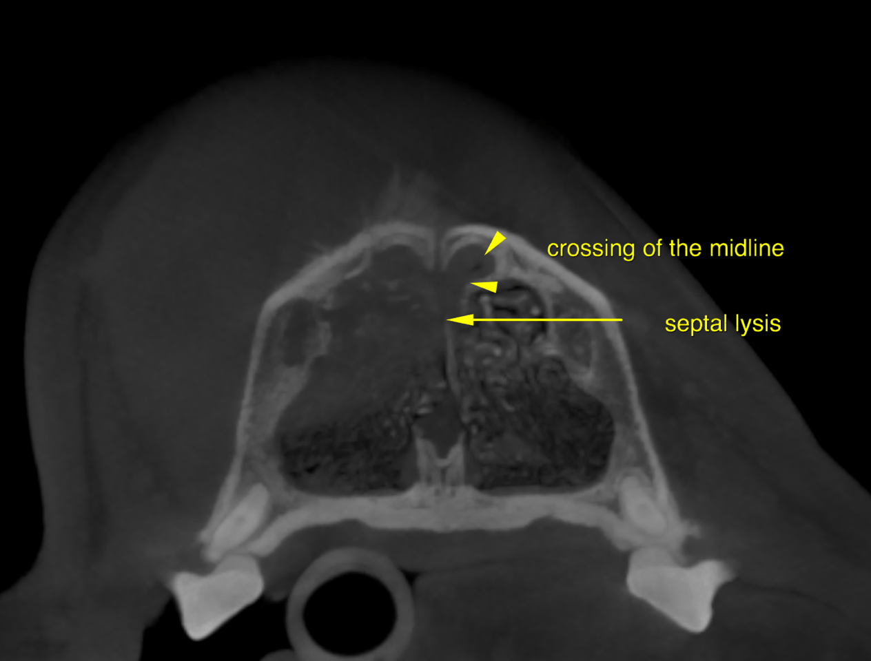
Triadan 104. There is focal lysis of the bony nasal septum with invasion of the left
nasal cavity noted.
The maxillary and nasal cavity extent of the mass spans the region from Triadan 104 to
108 (including these teeth).
After intravenous administration of iodinated contrast moderate, non-uniform
enhancement with predominanty peripheral distribution is noted within the mass.
The lymph nodes of the head are not included in the study.
Squamous cell carcinoma, lymphosarcoma, fibrosarcoma, hemangiosarcoma,
melanoma and mast cell tumor are differential diagnoses with a high potential.
Biopsy of the mass with consecutive histopathological examination is required for
further definition.
Therapeutic management is limited to palliative treatment options, as complete surgical
removal appears to be impossible even with aggressive hemimaxillectomy.
Radiation therapy and/or chemotherapy appear to be justifiable treatment options
depending on the tumor type.
If case this is an option for the owner, tumor staging should be completed including 3
view chest radiographs, ultrasound of the abdomen and fine needle aspiration of the
regional (mandibular & medial retropharyngeal) lymph nodes.