11_00060 Buffie H Umbilical hernia

Case Study

11_00060 Buffie H Umbilical hernia

A 9-year-old SF DSH was presented for evaluation of chronic anorexia and vomiting. Recently a lump had developed over the spay incision site – spay had been done a number of years ago.  Serum biochemistry, T4, and SDMA were all within reference range. Survey radiographs showed no obstructive pattern and slight swelling over the linea alba.
 

Sonographic Differential Diagnosis

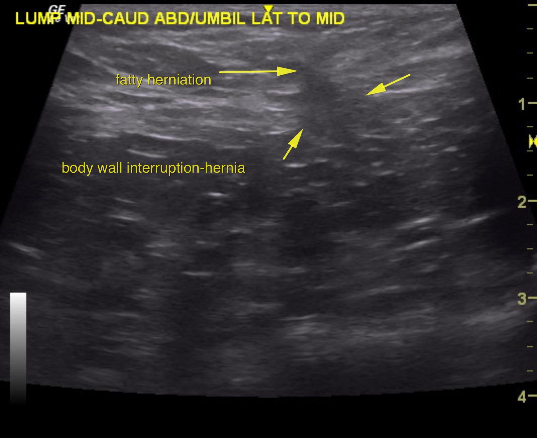
Hernia in the linea alba. Suspect umbilical 0.5 cm fatty extrusion. Mild potential for more pathological tissue. Recommend surgical correction of the hernia and given the age of the patient, biopsy of this tissue is always a good idea. Structurally the gastrointestinal tract is unremarkable. Treatment for food intolerance, possible helicobacter involvement should also be addressed.

Image Interpretation

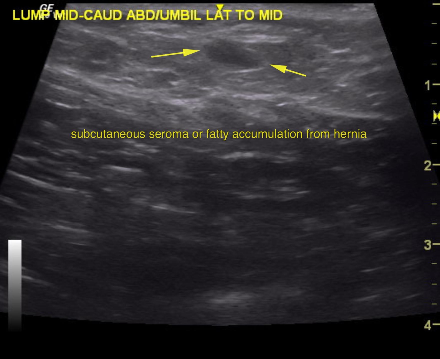
The swelling at the linea alba in this patient revealed a slight herniation of the body wall. Slight amount of fat accumulation was noted. The hernia measured approximately 0.5 cm. This is likely fatty extrusion; however, this tissue should be biopsied once surgically rectified. The ostium or hernia measures approximately 0.5 cm. No significant organ such as intestine are in the vicinity of the herniation. It appears that a 0.5 x 0.3 cm seroma formation or focal fatty accumulation is noted in the subcutaneous area which is causing the swelling.

DX

Umbilical hernia

Outcome

None

Clinical Differential Diagnosis

Lump – hernia, neoplasia, granuloma, abscess
Anorexia/vomiting – GI disease (IBD, food hypersensitivity, neoplasia, ulcer, foreign body); pancreatic disease (pancreatitis, neoplasia)

Sampling

None

Patient Information

Gender : Female, Spayed
Species : Feline
Type of Imaging : Ultrasound
Status : Complete

Clinical Signs

  • Anorexia
  • Vomiting

Images

as1as2

Clinical Signs

  • Anorexia
  • Vomiting