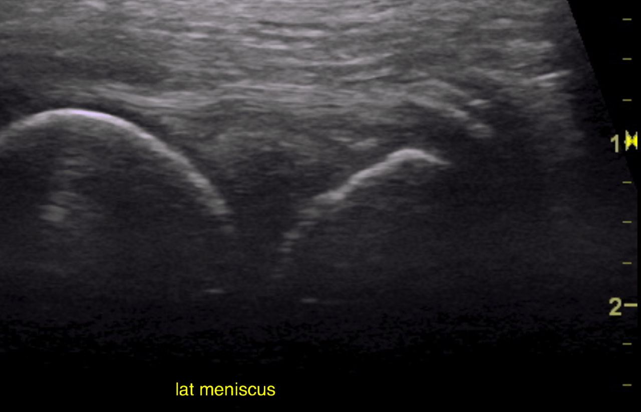
The patient is a Labrador Retriever mixed dog who has a history of a bilateral CCL injury. TTA surgery was performed on the left stifle July 2015. 2nd TTA on the right stifle is planned.
Physical exam: Positive cranial drawer, CTT, Medial buttress, no clicking
CBC/Chem wnl
The patient is a Labrador Retriever mixed dog who has a history of a bilateral CCL injury. TTA surgery was performed on the left stifle July 2015. 2nd TTA on the right stifle is planned.
Physical exam: Positive cranial drawer, CTT, Medial buttress, no clicking
CBC/Chem wnl