A 9-year-old NM Samoyed was presented for evaluation of recurrent hematuria, low SG, and occasional an abnormal urine stream. On urinalysis, SG 1.018 and 2+ blood was presented. Serum biochemistry was normal.
A 9-year-old NM Samoyed was presented for evaluation of recurrent hematuria, low SG, and occasional an abnormal urine stream. On urinalysis, SG 1.018 and 2+ blood was presented. Serum biochemistry was normal.
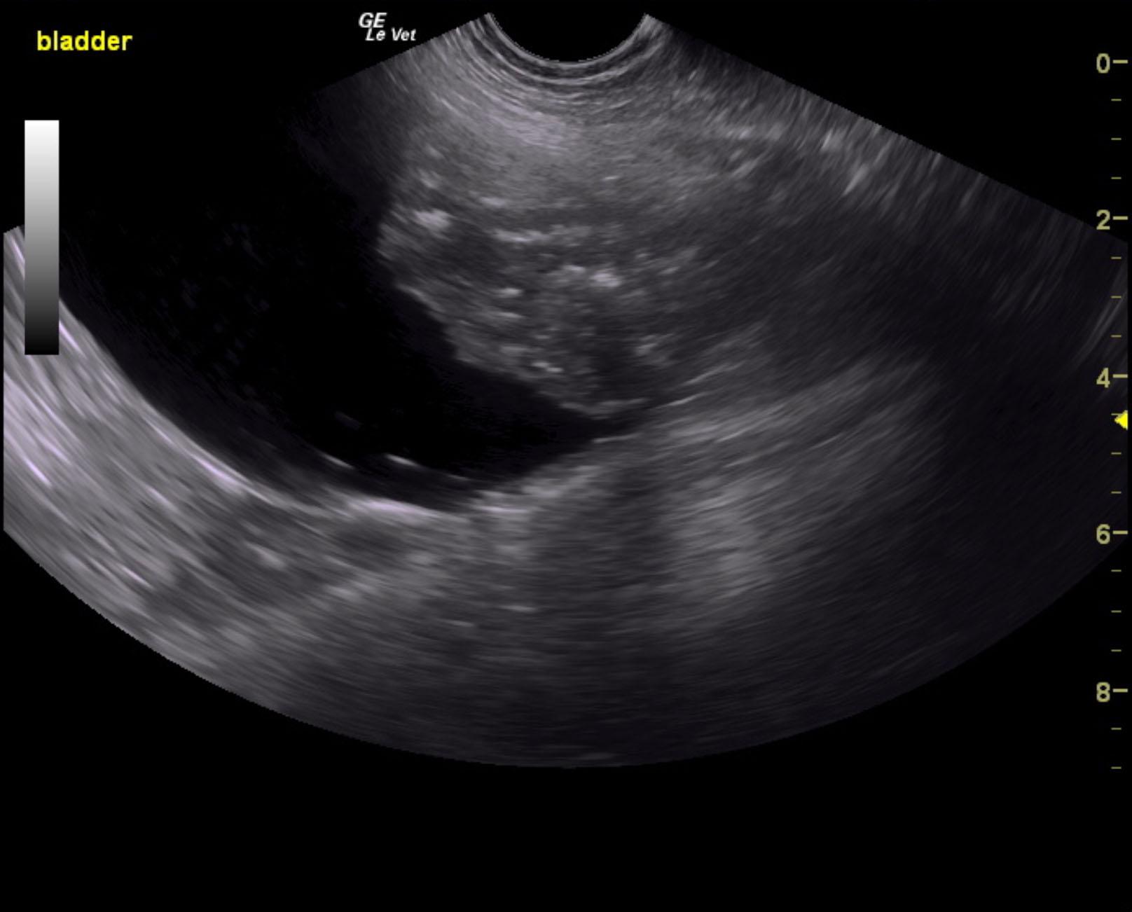
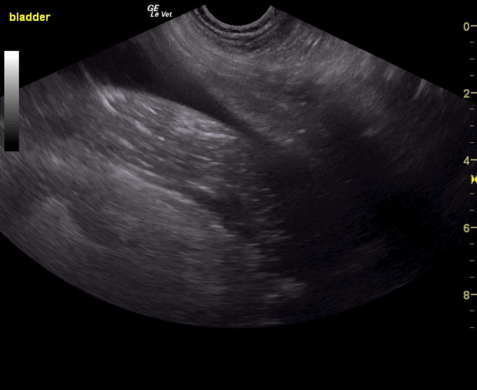
Bladder mass. Potentially resectable. This was encroaching on the cystourethral junction.
The urinary bladder presented a 6.0 cm, mixed, echogenic, mineralized mass that occupied the cystourethral junction and ventral bladder. The pelvic urethra was visible without overt invasion. However, the mass will act like a check valve upon the cystourethral junction. This extends to position of about 1.0 cm cranial to the cystourethral junction of the ventral wall. Aggressive resection may be possible. Vascularity was significantly high on power Doppler and color flow Doppler.
None
Prostate – neoplasia, prostatitis, abscess
Bladder – neoplasia, uroliths, bacterial cystitis, polyploid cystitis
Urethra – neoplasia, urethritis, lith
None