Case Study
RADS – Aspiration Pneumonia and Spondylosis Deformans in a 12 year old Cocker mix dog
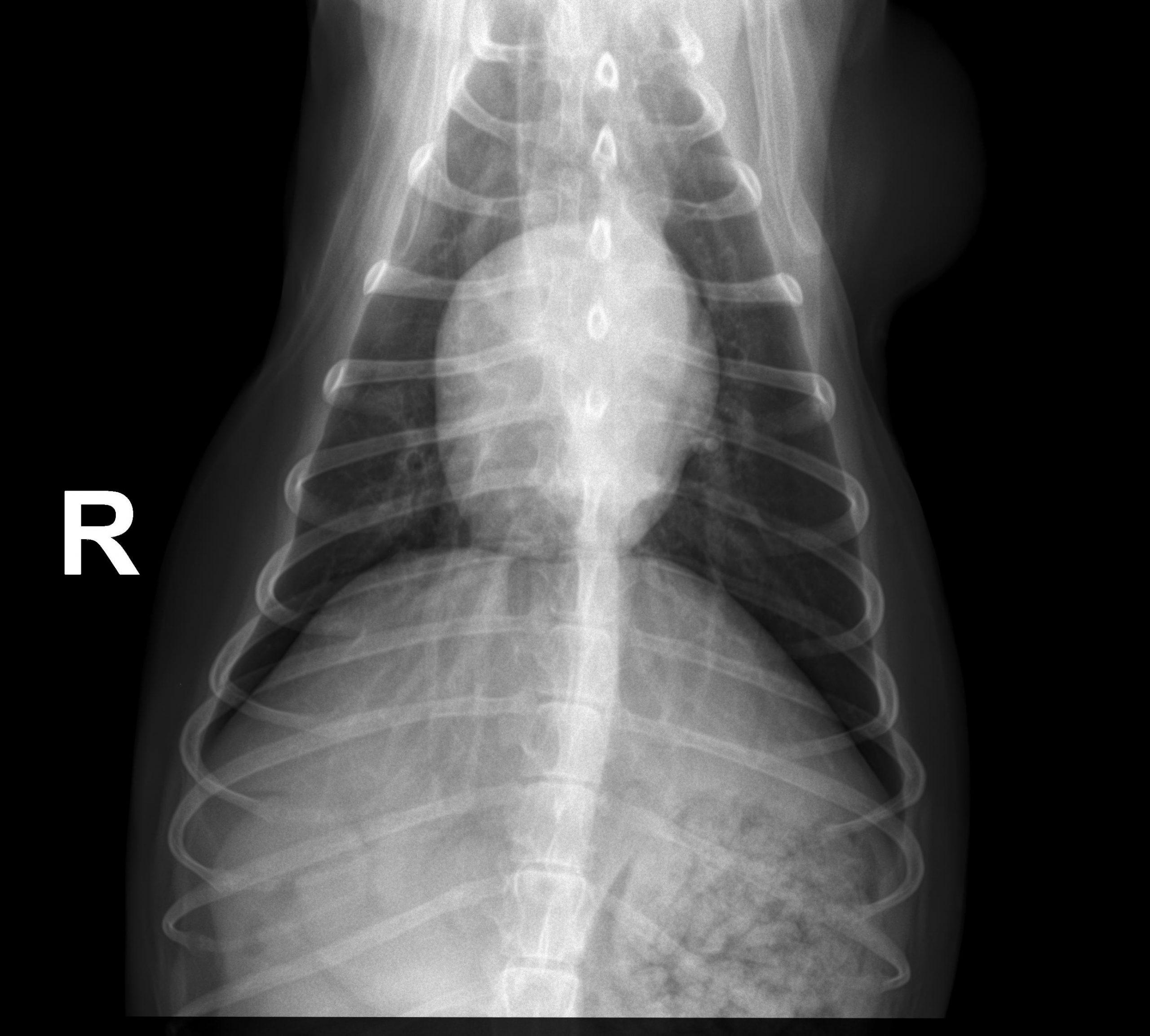
Image Interpretation
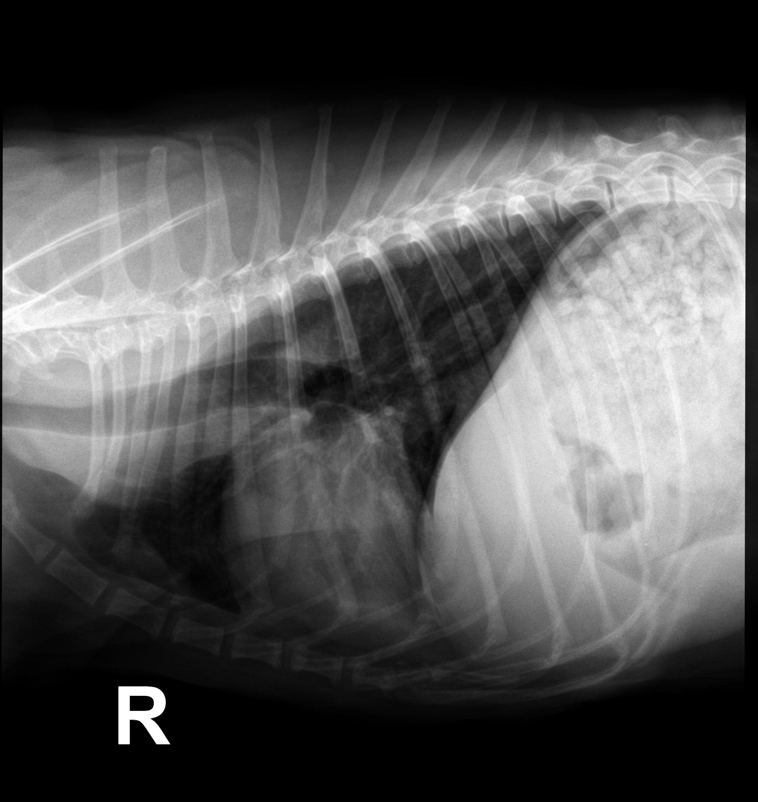
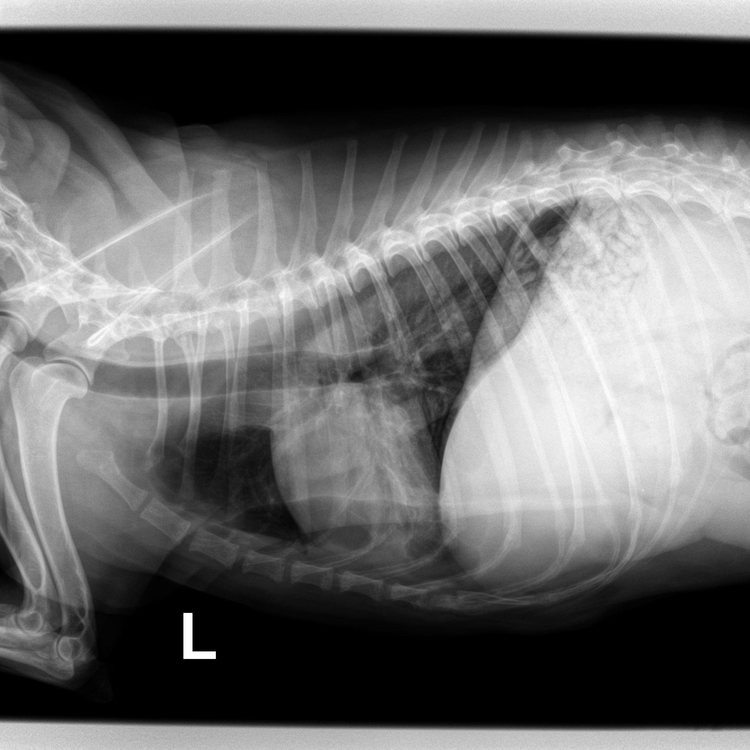
Rads of the thorax – There is a ventrally distributed alveolar pulmonary infiltrate involving the right middle
lung. Cardiovascular, pleural, lymphatic, and cranial mediastinal structures are normal.
The trachea diameter is normal. There is a small volume of air in the thoracic
esophagus. No rib or vertebral lesions are noted. The included thoracic limbs are
normal. There is an incidental finding of a large subcutaneous lipoma lateral to the left
shoulder on the VD view.
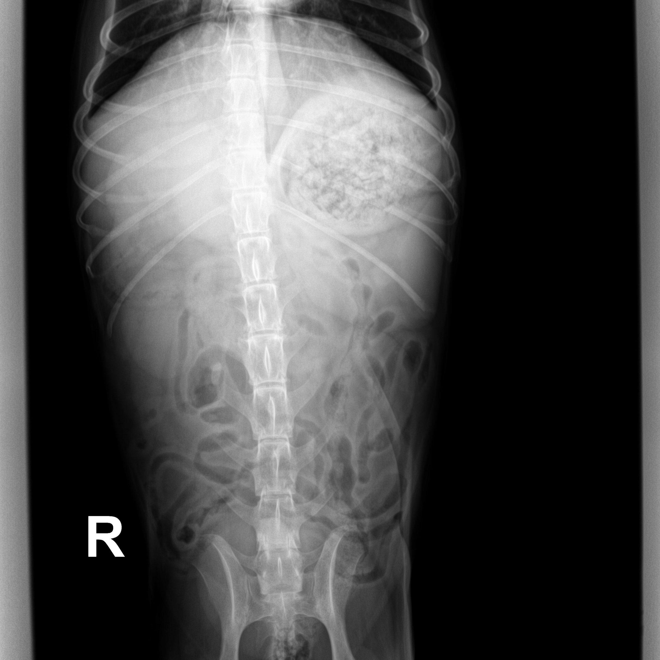
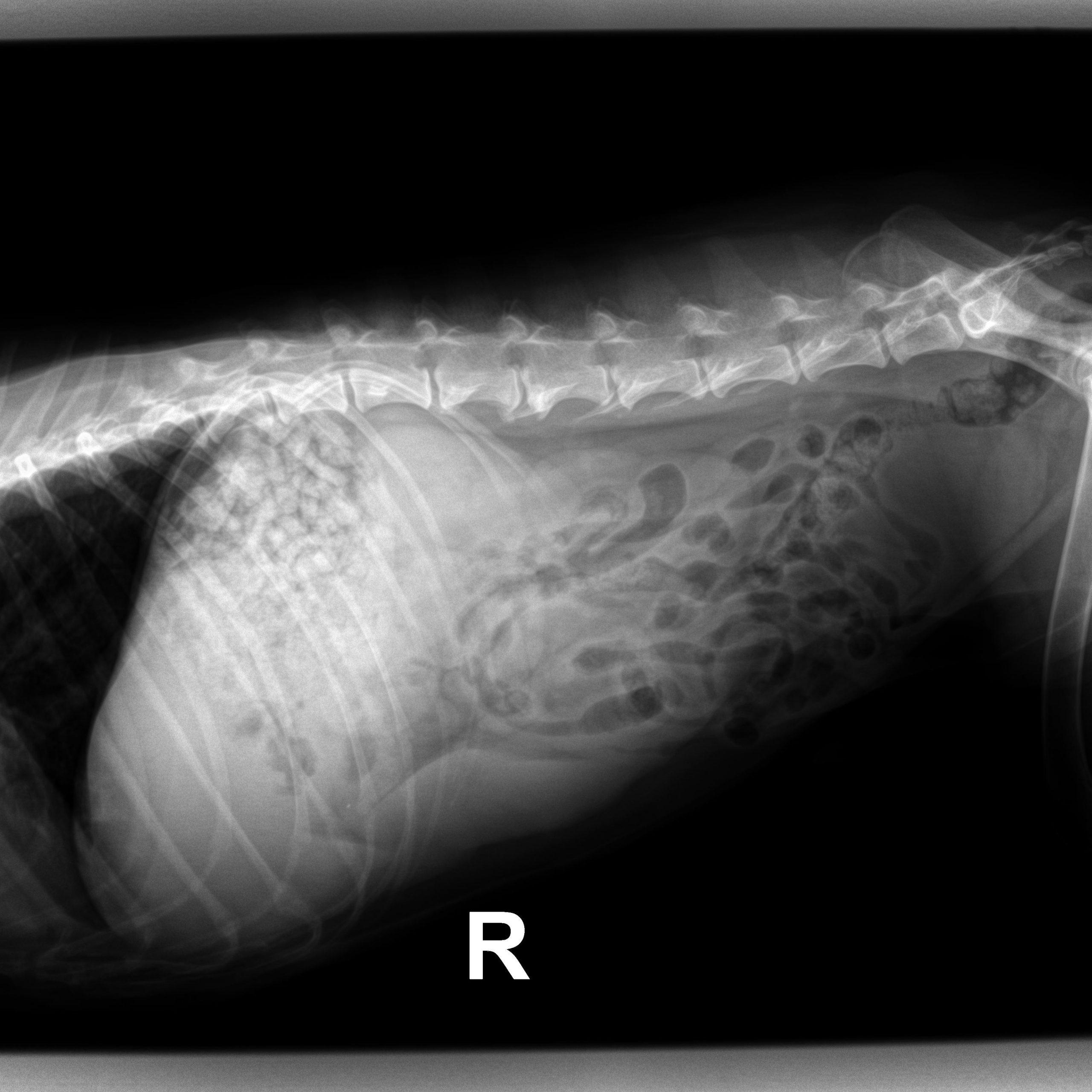
Rads of the abdomen – There is good abdominal detail. There is normal food material in the stomach. The
small intestines are empty, normal for diameter and distribution. There is a small
volume of fecal material in the colon. The liver and spleen are normal for volume and
margination. The urinary bladder, retroperitoneal space, kidneys, and area of the
pancreas are radiographically normal. There is mild L2-L3 spondylosis deformans.
Extra-abdominal skeletal structures are otherwise normal.
DX
Outcome
Recommendations:
1) Broad-spectrum antibiotic therapy is recommended.
2) The level of supportive care should be determined by clinical impressions and
physical examination findings.
3) Screening blood work is indicated, particularly to evaluate white blood count.
4) Repeat thoracic radiographs in 3–5 days to monitor response to therapy.
5) If there is clinical concern for laryngeal paralysis (could explain previous episode of
dyspnea as well as an underlying cause for seemingly random aspiration pneumonia),
direct evaluation of the larynx is indicated.
Patient Information
Clinical Signs
- Dyspnea
History
- Dyspnea
Images
Clinical Signs
- Dyspnea