A 2-year-old SF Maltese was presented for evaluation of hematuria, non-resolving UTI, and possibly PU/PD. CBC and serum biochemistry were within reference range.
A 2-year-old SF Maltese was presented for evaluation of hematuria, non-resolving UTI, and possibly PU/PD. CBC and serum biochemistry were within reference range.
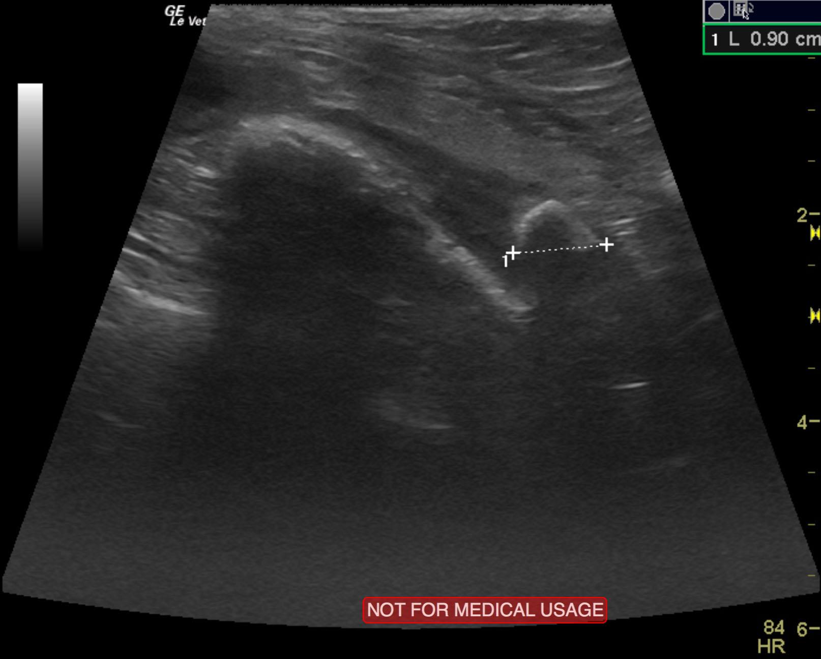
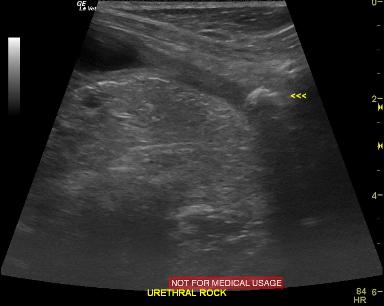
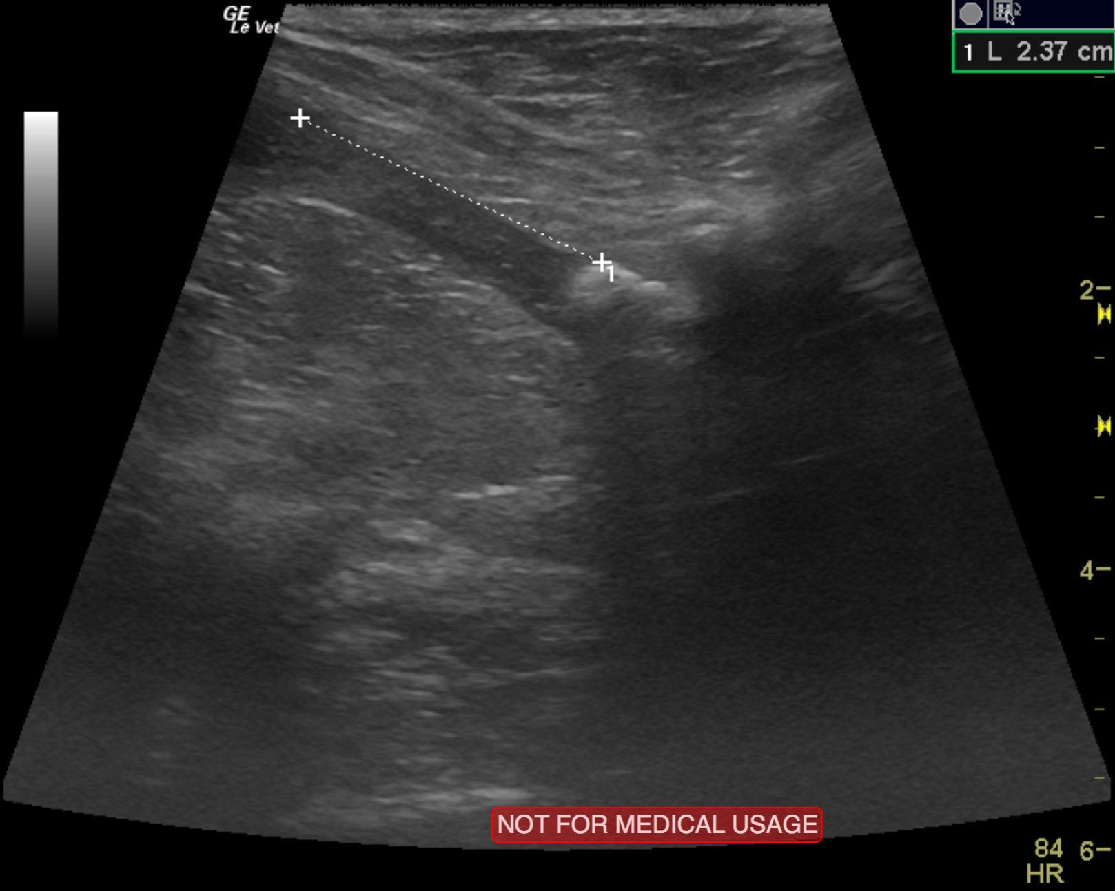
Large, embedded urethral calculus.
Concurrent urethritis.
Minor, regional free fluid. This is likely owing to inflammation, potential for urethral perforation.
The urinary bladder in this patient presented a pelvic urethral calculus that measured 0.9 cm with irregular edges embedded within the deep pelvic urethra and measured approximately 2.5 cm caudal to the cystourethral junction. Periserosal inflammatory pattern was noted along with a slight amount of free fluid noted adjacent to the urethra and calculus. The post calculus urethra appeared unremarkable. It appears that the passage of the calculus would entail approximately 1.5 cm of distal urethra in order to be evacuated. The bladder itself appeared unremarkable otherwise with a minor amount of debris.
None
Urinary bladder – chronic bacterial cystitis, urolith, persistent urachus, polyploid cystitis
Urethra – urolith, granulomatous urethritis
Kidney – renolith, pyelonephritis
None