CT Lumbosacral Disc Protrusion and Spondylosis with Pelvic Bladder in a German Shepherd Dog

Case Study

CT Lumbosacral Disc Protrusion and Spondylosis with Pelvic Bladder in a German Shepherd Dog


During a routine exam at the rDVM 6 weeks ago it was determined that the patient had lost weight. There is a long history of osteoarthritis and urinary incontinence.The recommendation was to increase food intake from 3 cups per day to 4 cups per day for a few weeks to see if the weight would return to normal (about 74 pounds). Upon recheck a few weeks later the weight had stabilized but not returned to normal.  Bloodwork showed elevated protein.  Full spinal radiographs were done, followed by referral for CT. Fungal serology on 6/25 was negative. The patient is eating well and drinking more than normal, unsure whether clincial issue or due to warmer weather.  No V/D. No history of seizures or allergies. Medications include Proin 25mg BID, no vitamins or supplements. 

Exam Findings: Weight: 70.8 pounds (7/10/2015) Pulse: 90; Respiration Rate: pant; CRT: 2 MM; Color: P/M, hydrated 

Temperature: not taken due to owner request.  BAR, with a body condition score of 5/9. Eyes, ears, nose and throat appeared normal. A moderate amount of dental calculus was noted on the teeth, and the canine teeth were very worn. The coat was full with no evidence of ectoparasites. On auscultation the heart and lungs sounded normal with no murmurs or arrhythmias noted. Abdominal palpation was within normal limits with no masses or organomegaly noted. Neurologic system appeared to be normal. The gait was normal at a walk and trot today. Musculoskeletal system appeared to be within normal limits. There were no abnormalities noted on orthopedic examination. Pain Scale (Colorado 0-4) – 1/4 (slightly painful with pressure on lumbar spine.
DX

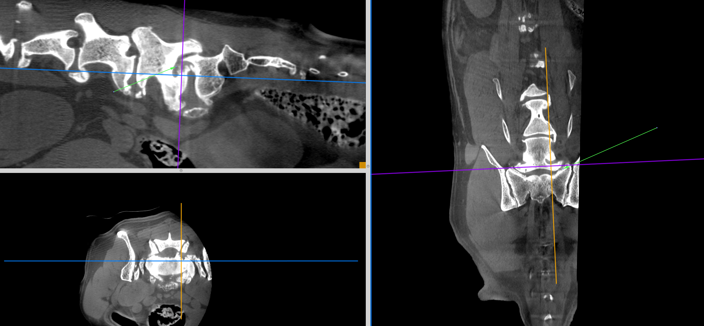
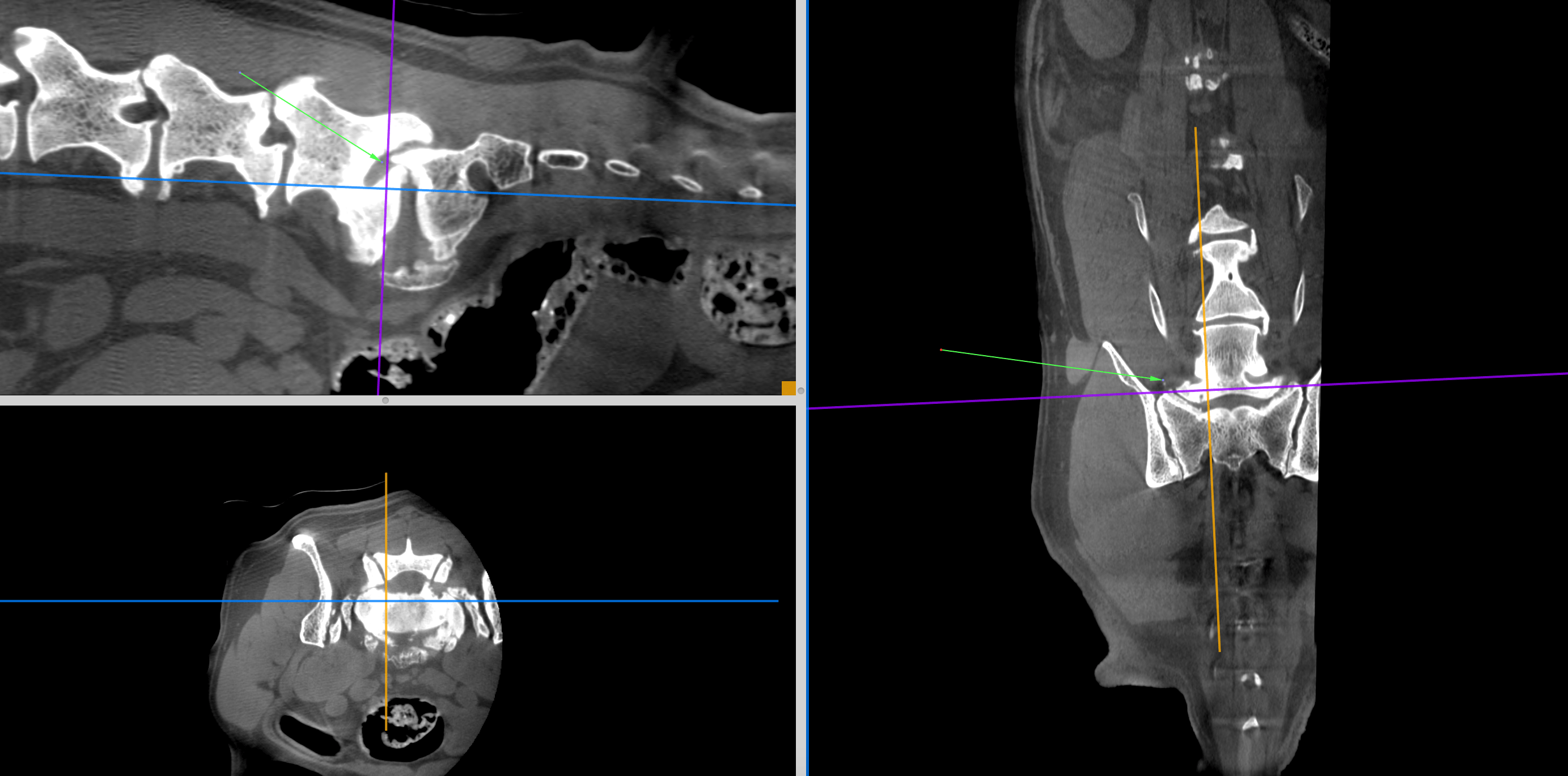
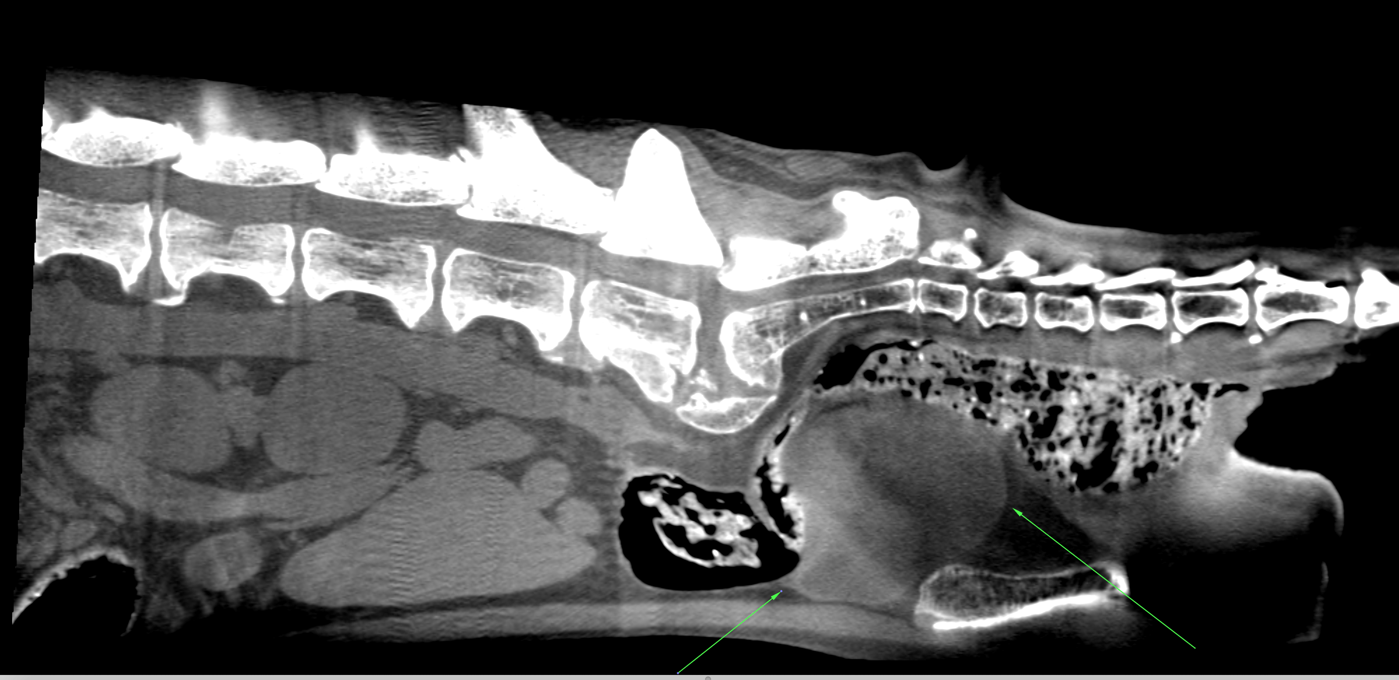
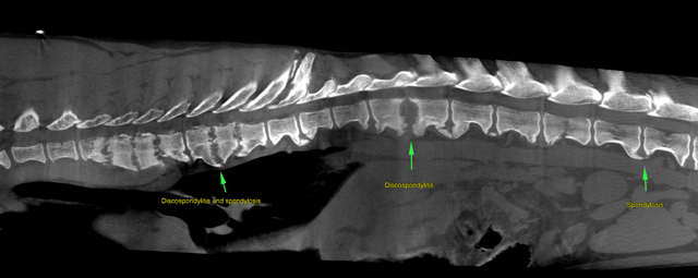
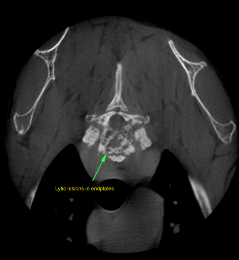
An aggressive osteolytic process was not seen. There was degenerative lumbosacral and neuroforaminal stenosis due to severe lumbosacral disc protrusion and spondylosis resulting associated with compressive neuropathy of the Cauda equina nerve roots level with the vertebral canal and neuroforamina (L>>R). Mild chronic disc protrusions with generalized degenerative changes of the intervertebral connections were present throughout the lumbar spine. The clinical relevance of these changes is questionable as there was no compressive myelopathy. There was mild right sided coxarthrosis and moderate bilateral iliosacral joint arthritis, both of which are likely unrelated to the clinical signs. There was a pelvic bladder which may explain the urinary incontinence. There was splenic congestion which likely was a function of the anesthetic protocol and/or German Shepherd Dog hypersplenism.

Image Interpretation

CT of the lumbar spine: The scan covers lumbar spine from L3 caudally and the lumbosacral transition. The thoracic spine and a post contrast series were not available for assessment. No signs of an aggressive osteolytic process were noted. There was severe protrusion of the lumbosacral disc, severe lumbosacral spondylosis, mild step formation and telescoping of the sacral vertebral lamina. The epidural fat signal and Cauda equina nerve roots were not discernible. There was bilateral foraminal stenosis throughout the entrance and exit zones which was moderate on the right and severe on the left side. The foraminal stenosis was mainly due to lateral and dorsal proliferation of the lumbosacral spondylosis. The left and right nerve roots of L7 were not seen within their neuroforamina.
Mild intervertebral disc protrusions without significant compressive myelopathy were seen throughout the lumbar spine from L3 to L7. There was also mild to moderate spondylosis and spondylarthrosis throughout the lumbar spine as imaged.
There was caudal lumbar dural calcification.
Both iliosacral joints showed moderate arthrotic changes.
The right hip joint demonstrated mild osteoarthrosis.
The urinary bladder was in a caudal, partially intrapelvic position.
There was moderate splenic enlargement.

Patient Information

Patient Name : Ruby Farnady, Sierra Veterinary Specialists
Gender : Female, Spayed
Species : Canine
Type of Imaging : Ultrasound

Clinical Signs

  • Weight loss

History

  • Incontinence

Images

screen_shot_2015-07-24_at_10screen_shot_2015-07-24_at_10bildschirmfoto_2015-07-24_um_00bildschirmfoto_2015-07-24_um_00bildschirmfoto_2015-07-24_um_00

Blood Chemistry

  • Total Protein, High

Clinical Signs

  • Weight loss
Skip to content