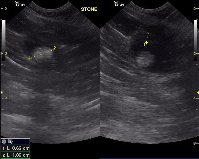
An 8-year-old female DSH was presented for evaluation of hematuria.
Sonographic Differential Diagnosis
Interstitial cystitis with concurrent bladder stone.
A cystotomy, bladder wall biopsy and culture, and stone analysis are recommended. Part of the underlying issue may not be only the calculus but also urinary bladder wall disease.
Image Interpretation
A 1.1 cm uniform, shadowing calculus was noted in the urinary bladder. However, the bladder wall itself revealed a 0.6 cm ventral apical wall thickening consistent with interstitial cystitis or similar.
DX
Chronic cystitis with hemorrhage
Outcome
Cystotomy was performed and revealed struvite stone. The patient was placed on C/D diet. The patient is doing well at home.