06-00243 Rylee N Bilat ureteroliths—— NEED CDX——-

Case Study

06-00243 Rylee N Bilat ureteroliths—— NEED CDX——-

A 9 year-old SF canine Shih Tzu was presented one month ago for severe hematuria and not acting herself. Nine days prior to initial presentation blood work demonstrated BUN 44. Phosphorus 10.6. Creatinine 4.6. Glucose 113. UA: Specific gravity 1.020 with a large amount of blood and 4+ proteinuria. Ultrasound at that time showed ureterolithiasis and a dilated ureter on the left. Bladder calculi and pyelectasia were noted in both kidneys. At that time the right kidney revealed a 0.35 cm calculus at the corticomedullary junction and pyelectasia that measured 0.72 cm.

A 9 year-old SF canine Shih Tzu was presented one month ago for severe hematuria and not acting herself. Nine days prior to initial presentation blood work demonstrated BUN 44. Phosphorus 10.6. Creatinine 4.6. Glucose 113. UA: Specific gravity 1.020 with a large amount of blood and 4+ proteinuria. Ultrasound at that time showed ureterolithiasis and a dilated ureter on the left. Bladder calculi and pyelectasia were noted in both kidneys. At that time the right kidney revealed a 0.35 cm calculus at the corticomedullary junction and pyelectasia that measured 0.72 cm. The right kidney measured 3.75 cm. Right ureteral calculus was embedded in the right renal pelvis and measured 1.5 cm. The left kidney revealed pyelectasia 0.28 cm and the left kidney measured 3.31 cm.  Left ureteral calculus measured 0.4 cm. Bladder calculi measured 0.1—0.3 cm. Rylee is feeling much better, eating drinking well, gained back some weight. Creatinine 1.5. urinalysis revealed marked bacteria, urine specific gravity 1.020, trace protein. BUN 44. The patient has been on one month of medical treatment.
 

Sonographic Differential Diagnosis

Persistent right ureteral calculi with persistent pyelectasia and ureteritis.
Some urine is likely passing in the right ureter given that the hydronephrosis of the right kidney has only progressed in a minor fashion. The left kidney has significantly improved. Surgical intervention is still recommended in this patient.

Image Interpretation

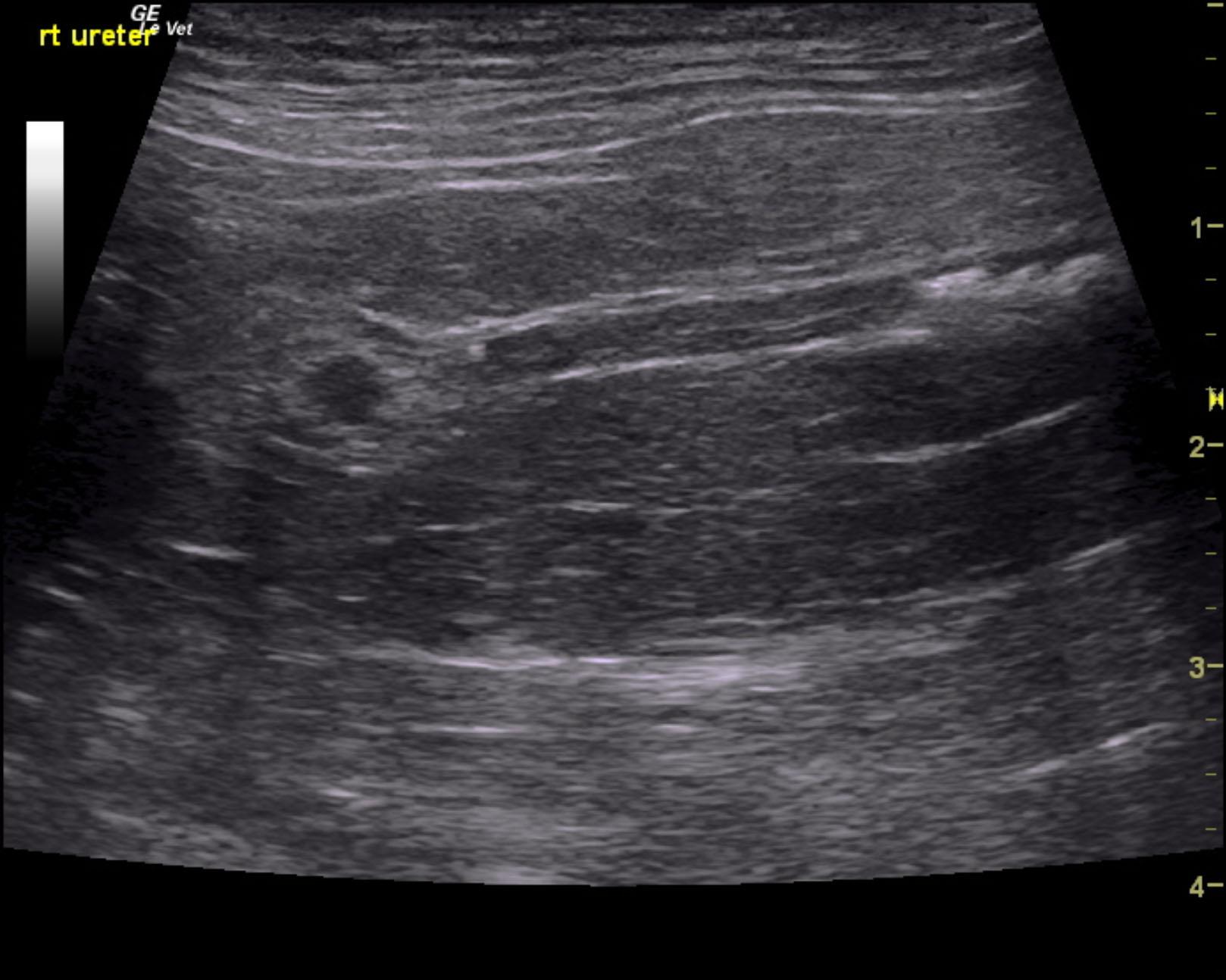
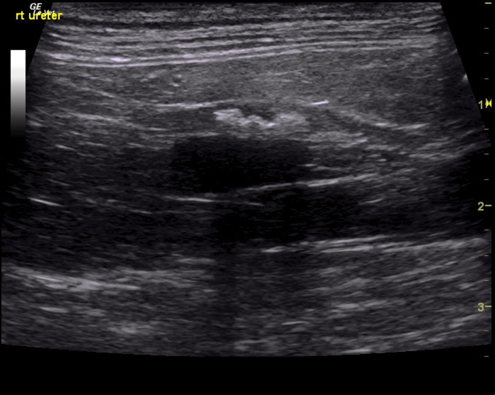
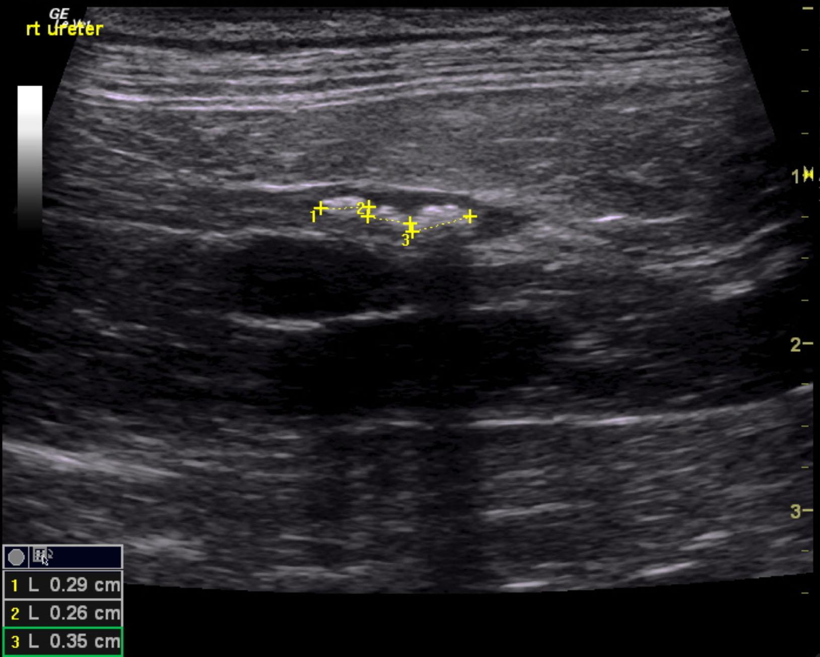
The urinary bladder was poorly distended at this time. A trace 0.2 cm, non obstructive bladder stone was noted. Three separate calculi were noted and measured 0.35 cm, 0.26 cm, and 0.29 cm in the distal right ureter. The ureter was thickened with echogenic debris. This is consistent with ureteritis with multiple embedded calculi. Pyelectasia was persistently present in the right kidney measuring 0.84 cm, which is mildly progressed. Calculus was persistently present in the right renal pelvis and measured 0.4 cm. Pyelectasia of the left kidney was persistent and measured 0.12 cm. The left kidney measured 3.09 cm with mild to moderate degenerative changes. The left kidney has significantly improved compared to the prior sonogram.

DX

Bilateral ureteroliths

Outcome

None

Comments

Need whole case. Two US, report from first, images from 2nd. Sep ’15 asked for the other report.

Sampling

None

UA Specific Gravity Range

1.020

Patient Information

Gender : Female, Spayed
Species : Canine
Type of Imaging : Ultrasound

Clinical Signs

  • "Not Doing Right"
  • Anorexia

Images

rieley_nemex_persistent_ureteral_stones_1_month_med_txrieley_nemex_persistent_ureteral_stones_1_month_med_tx_2rieley_nemex_persistent_ureteral_stones_1_month_med_tx_3

Blood Chemistry

  • BUN high
  • Creatinine, High

Clinical Signs

  • "Not Doing Right"
  • Anorexia

Urinalysi

  • Blood Present
  • Protein Present