The patient is a 10 year old MN Pug dog, which was presented for hematuria. Radiograph is unremarkable with the exception of some spondylosis. Abnormalities on blood work included an elevated ALP at 259 and a low BUN at 4.
The patient is a 10 year old MN Pug dog, which was presented for hematuria. Radiograph is unremarkable with the exception of some spondylosis. Abnormalities on blood work included an elevated ALP at 259 and a low BUN at 4.
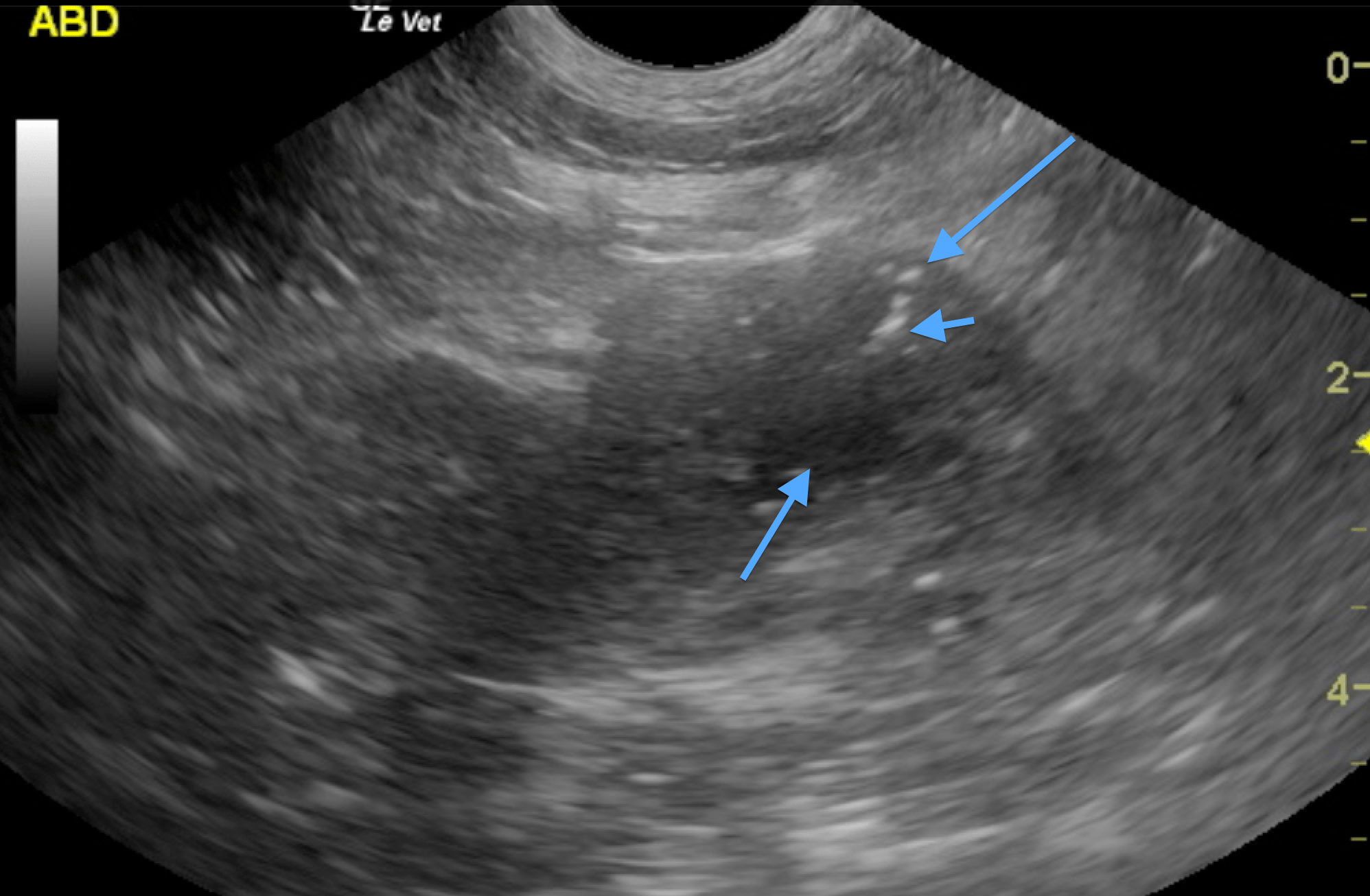
Prostatic mass strongly suspicious for prostatic carcinoma with regional inflammation. No overt evidence of metastatic disease noted; however chest radiographs would be recommended to rule out metastatic disease.
The urinary bladder was structurally unremarkable, however the prostate and pelvic urethra contained a mass measuring approximately 2.5 cm with mineralization and a pericapsular inflammatory pattern. The pelvic urethra appeared largely obliterated by the mass infiltration. Nodular hypoechoic changes also noted. A cystic portion of the prostate was also noted. The middle arrow indicates the prostatic cyst, while the small arrow indicates the presence of dystrophic mineralization. The iliac lymph nodes did not appear involved.
None
Urethral stent placement is an option in this patient could be performed at Animal Medical Center with Dr. Chick Weisse.
UTI, urinary prostatic neoplasia, urolithiasis, coagulopathy.
Ultrasound guided fine needle aspirates were performed without complication. The long arrow indicates the location of the FNA. Cytology revealed carcinoma.