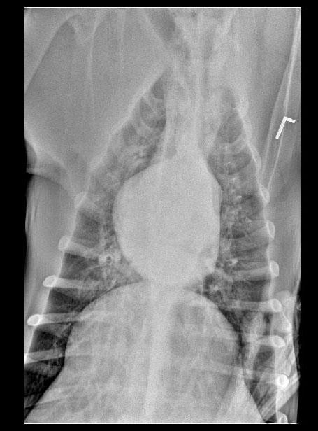
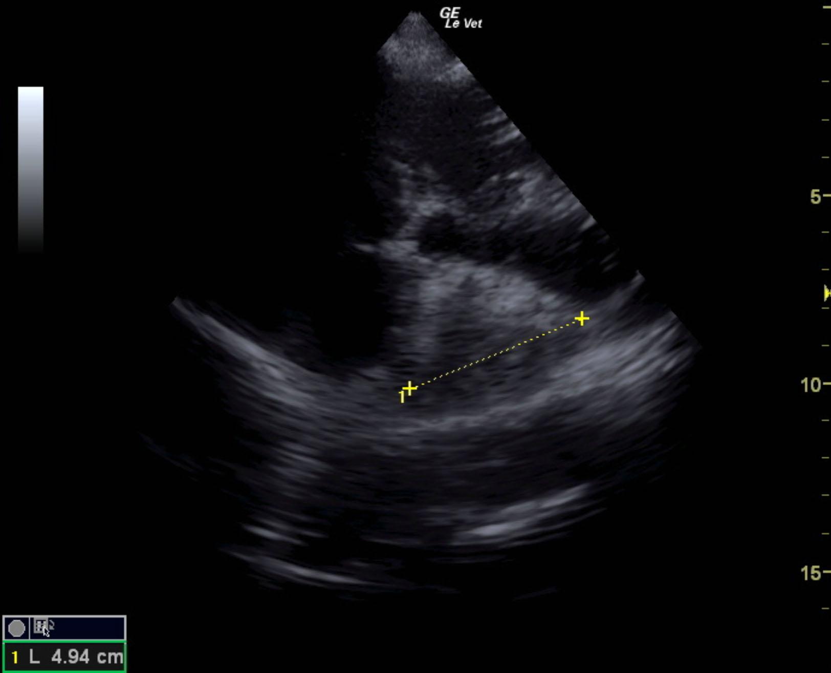
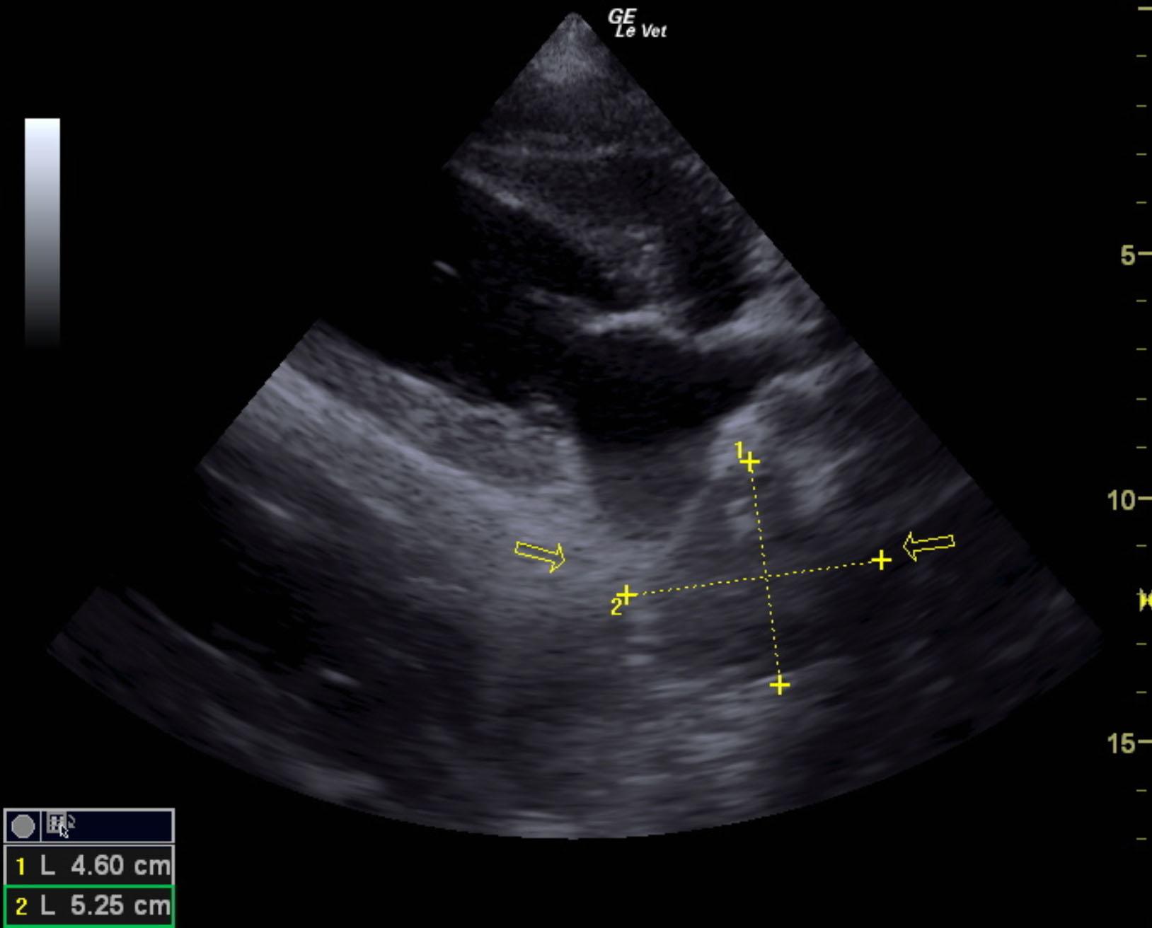
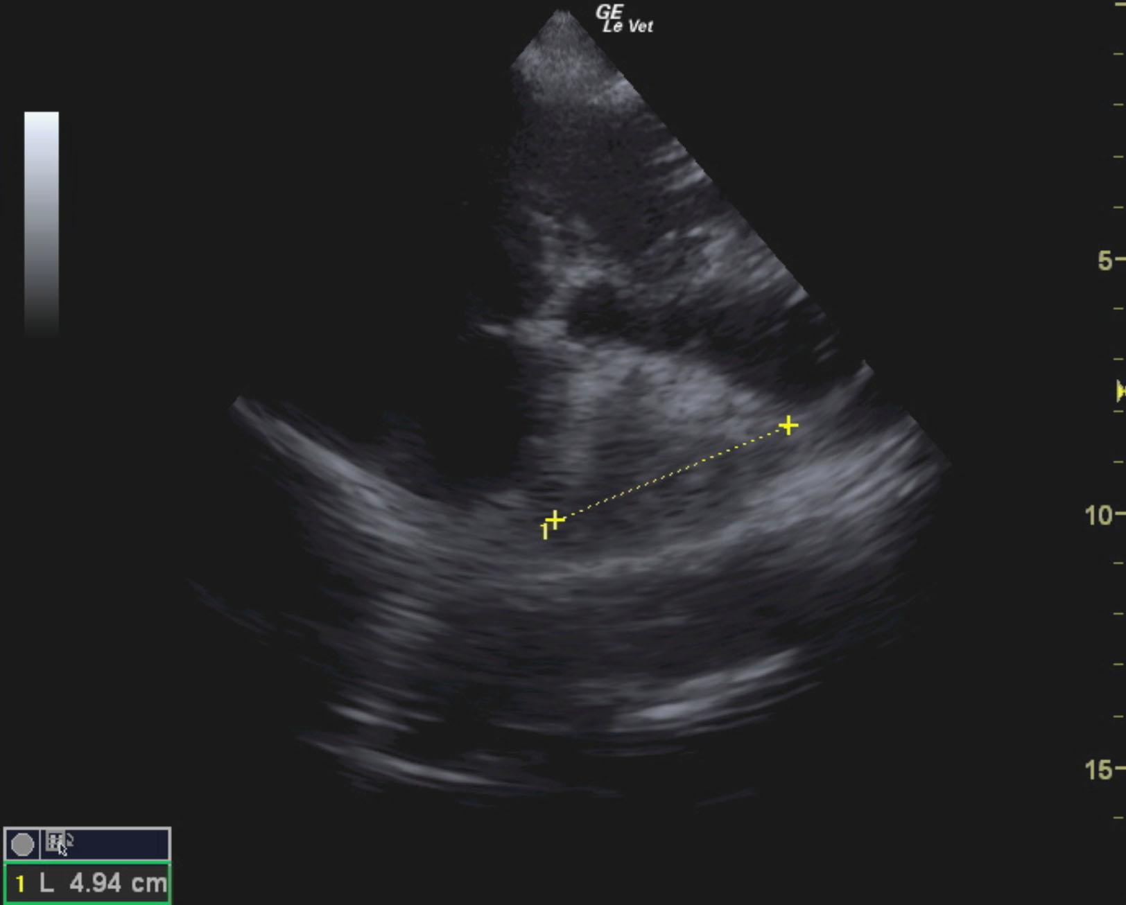
The patient is a canine Mastiff, male, 1 year old who was presented for surgery for the eye, however physical exam revealed muffled heart sounds. Radiographs revealed mass at the heart base.
The patient is a canine Mastiff, male, 1 year old who was presented for surgery for the eye, however physical exam revealed muffled heart sounds. Radiographs revealed mass at the heart base.