The patient is a 12-year-old MN Dachshund, who was presented for dribbling urine. The dog was unwilling or unable o urinate on leash. A urinary catheter passed easily. The rectal exam was unremarkable.
The patient is a 12-year-old MN Dachshund, who was presented for dribbling urine. The dog was unwilling or unable o urinate on leash. A urinary catheter passed easily. The rectal exam was unremarkable.
Case Study
Prostatic mass with bilateral ureteral obstruction in a 12 year old MN Dachshund
Sonographic Differential Diagnosis
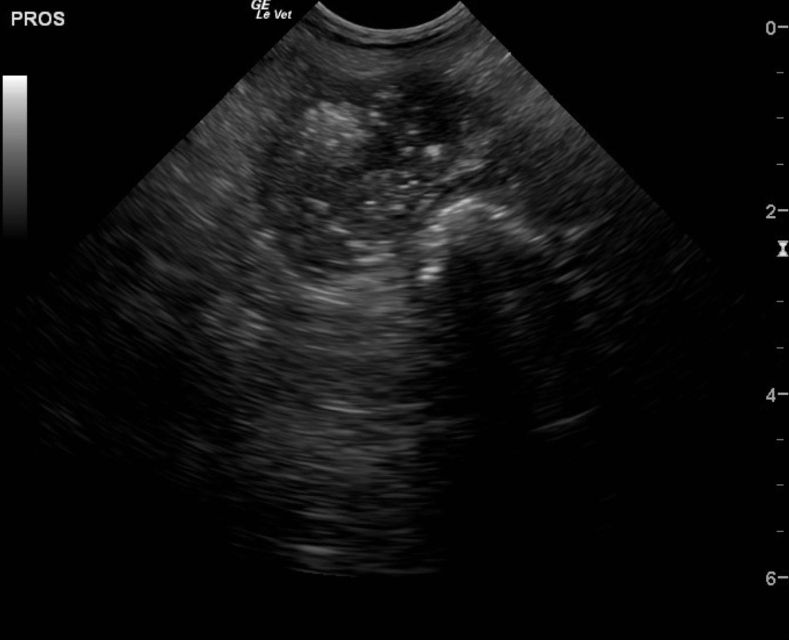
Prostatic mass with urethral involvement and obstruction. This is strongly suggestive of prostatic carcinoma. Secondary bilateral ureteral dilation. Mild hydronephrosis due to chronic increased urinary pressures.
Image Interpretation
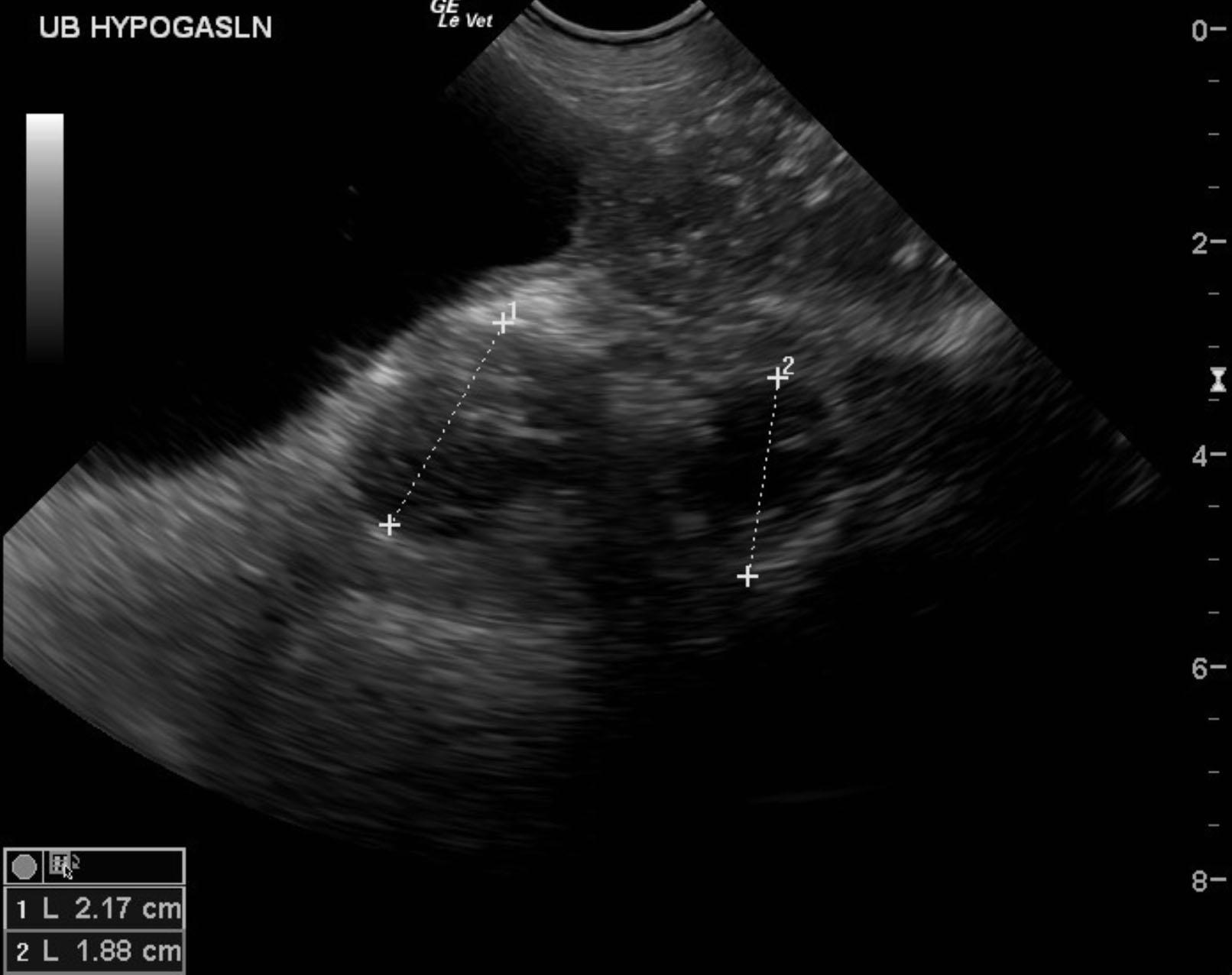
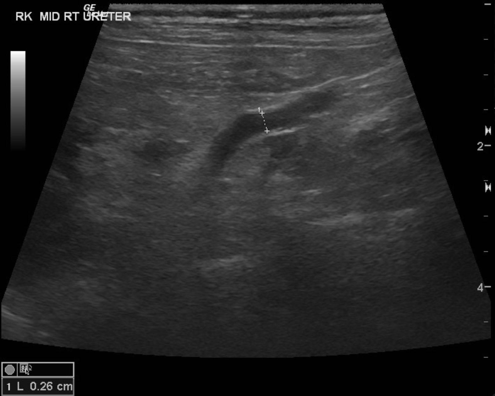
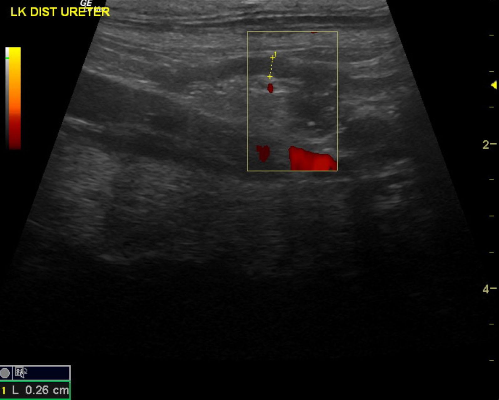
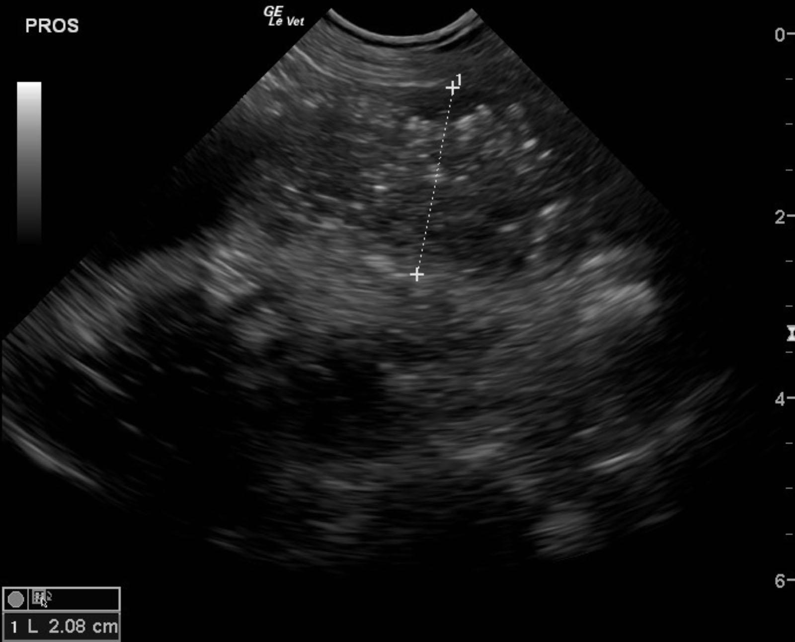
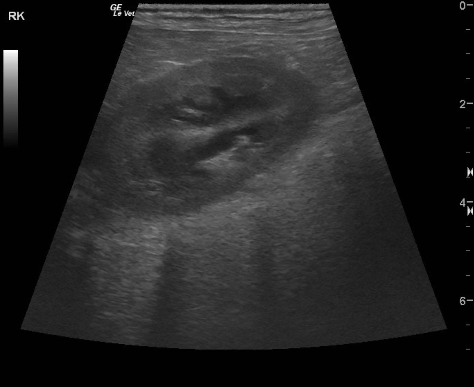
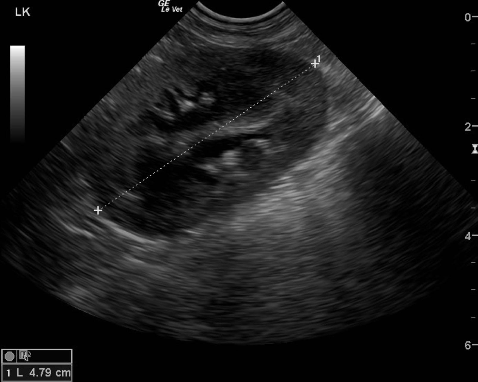
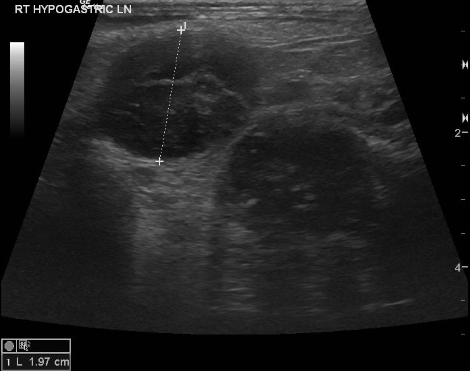
The urinary bladder and trigone appeared normal. The right ureter was dilated and tortuous measuring 0.32 cm. The left ureter was also mildly dilated at 0.26 cm. The ureteral dilation is likely owing to stasis and urinary outflow issues due to urethral obstruction. No neoplastic invasion into the ureters was noted. The prostate was enlarged and hypoechoic with multi-focal, hyperechoic areas of mineralization and capsular expansion. The prostate measured 2.6 cm entering into the urethra and extended cranially to the cystourethral junction creating an obstructive pattern. The kidneys revealed minor degenerative changes and slight, dystrophic mineralization. Pyelectasia was noted in the right kidney at 1.0 cm and 1.33 cm in the left kidney. The left kidney was swollen at 4.8 cm. The right kidney measured approximately 4.5 cm. A hypogastric lymph node was enlarged, hypoechoic and irregular measuring 1.97 cm with other regional lymph nodes. These lymph nodes were also focally mineralized.
DX
Prostatic mass with bilateral ureteral obstruction
Outcome
None
Comments
Cox-2 inhibitor therapy may be added to the protocol +/- chemotherapy depending upon oncology consultation. However, urethral stent placement is strongly recommended.
Clinical Differential Diagnosis
Urinary incontinence, UTI, prostatic neoplasia, urinary neoplasia, urolithiasis.
Sampling
Ultrasound-guided FNA of the hypogastric lymph node and prostatic mass would be recommended, however, none was completed.
Video
Patient Information
Patient Name :
Bruno C
Gender :
Male, Neutered
Species :
Canine
Type of Imaging : Ultrasound
Status :
Complete
Liz Wuz Here :
Yes
Code :
09_00074
Clinical Signs
- Hematuria
- Incontinence
Images
Clinical Signs
- Hematuria
- Incontinence