The patient is an 8 year old neutered male canine Queensland Heeler mix dog that presented for hematuria. Clinical exam was unremarkable. CBC and chemistry panel were unremarkable. Hematuria and evidence of UTI were present in the urinalysis.
The patient is an 8 year old neutered male canine Queensland Heeler mix dog that presented for hematuria. Clinical exam was unremarkable. CBC and chemistry panel were unremarkable. Hematuria and evidence of UTI were present in the urinalysis.
Case Study
Ureteral sarcoma and hydronephrosis in an 8 year old MN Queensland Heeler mix with hematuria
Sonographic Differential Diagnosis
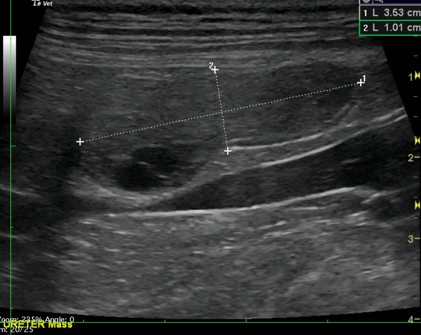
Obstructive right ureteral mass with extension into right renal pelvis and secondary hydronephrosis. Potential renal pelvic blood clot. Mass appears resectable with right nephroureterectomy. Suspect transitional cell carcinoma or sarcoma.
Image Interpretation
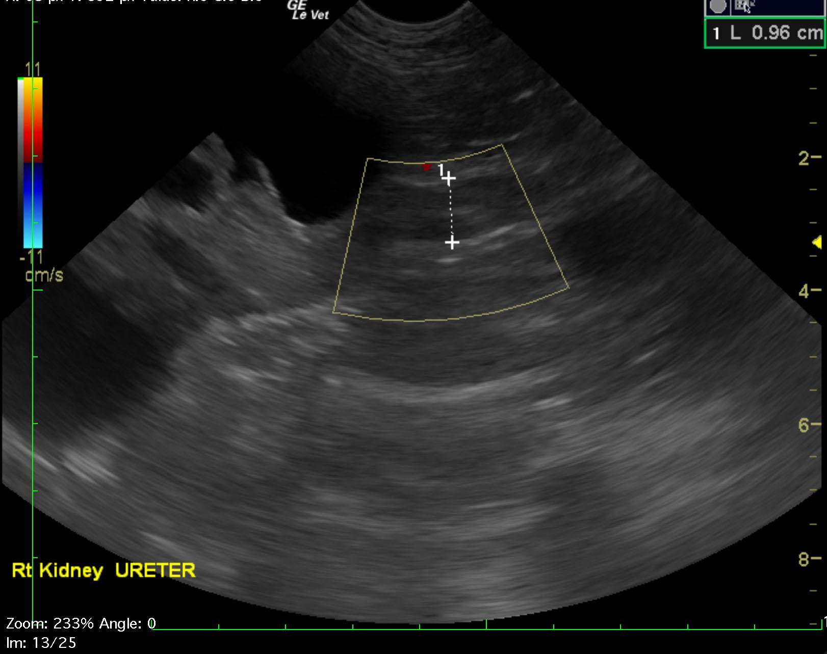
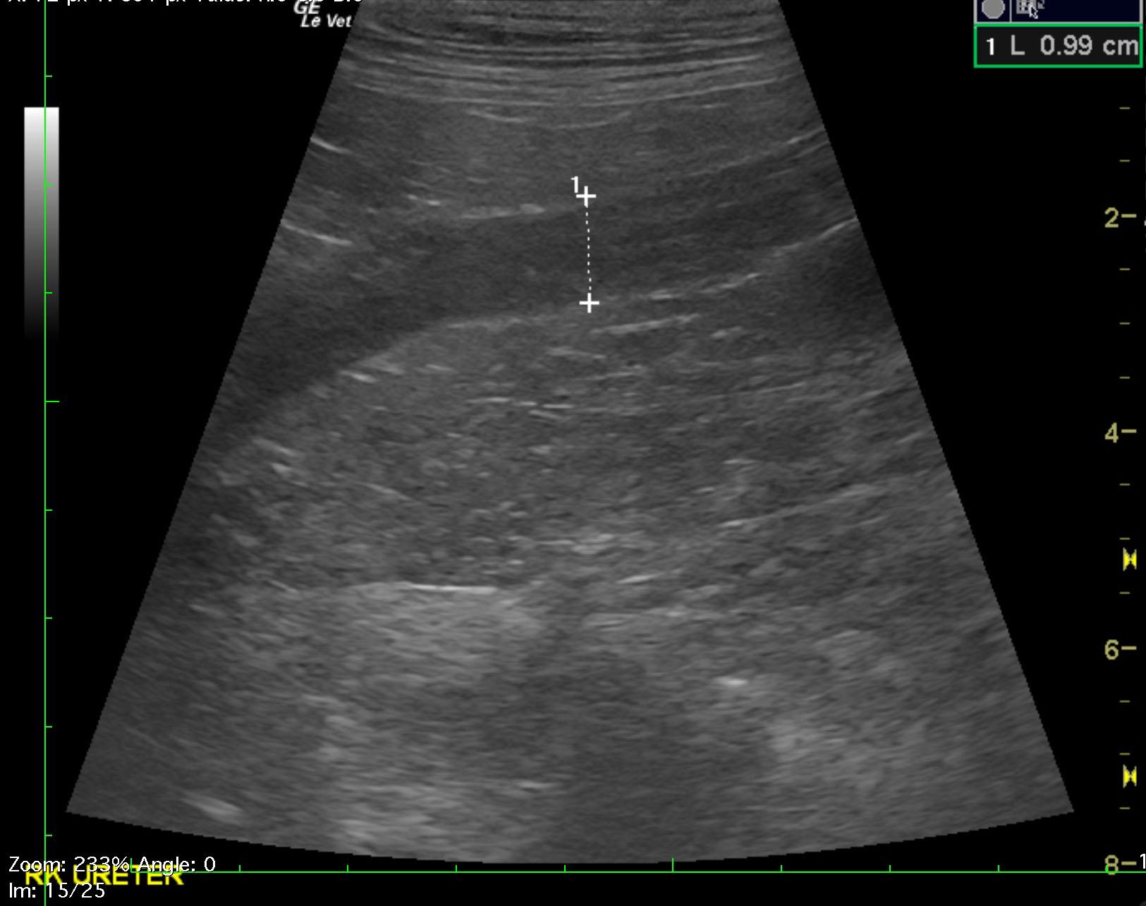
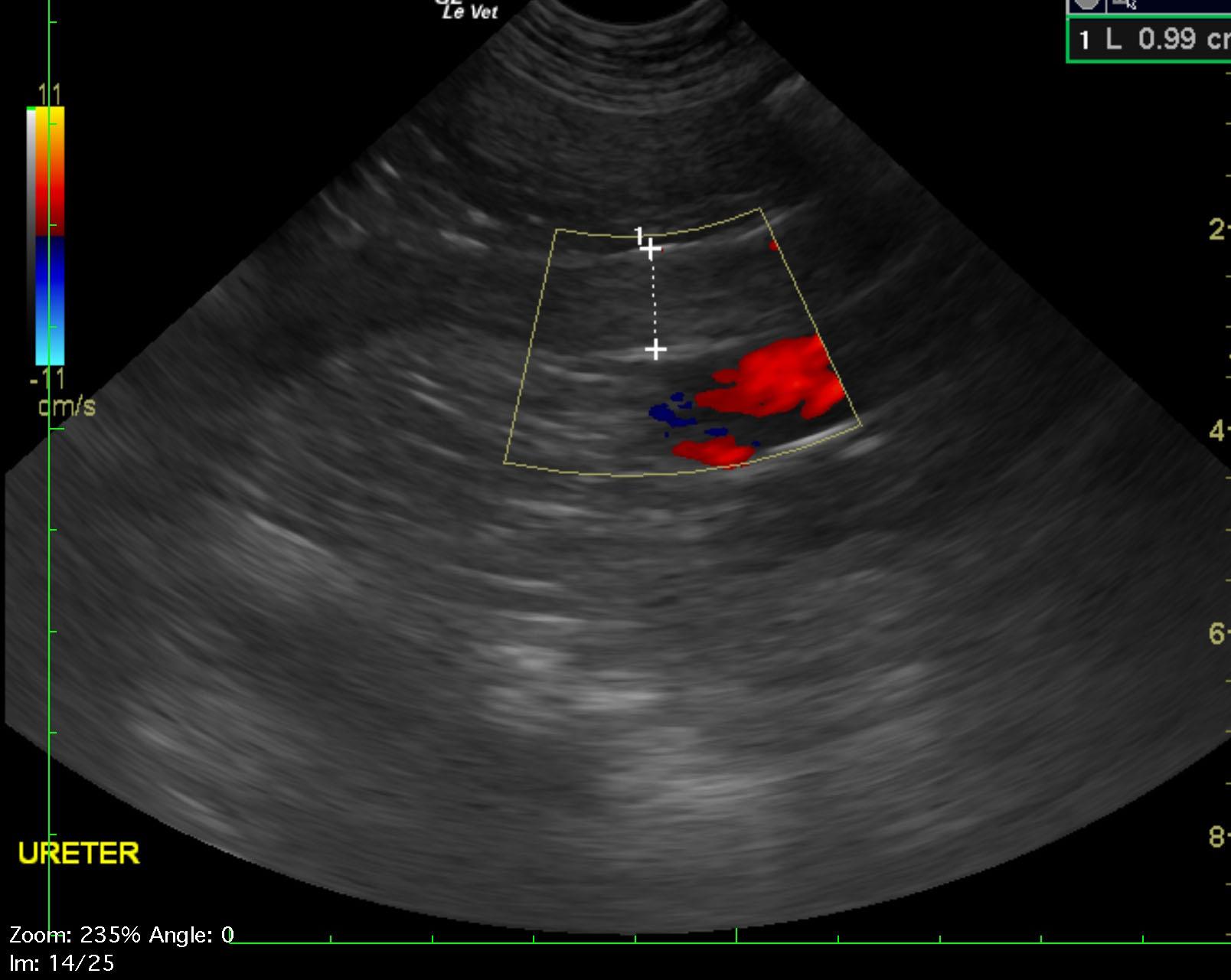
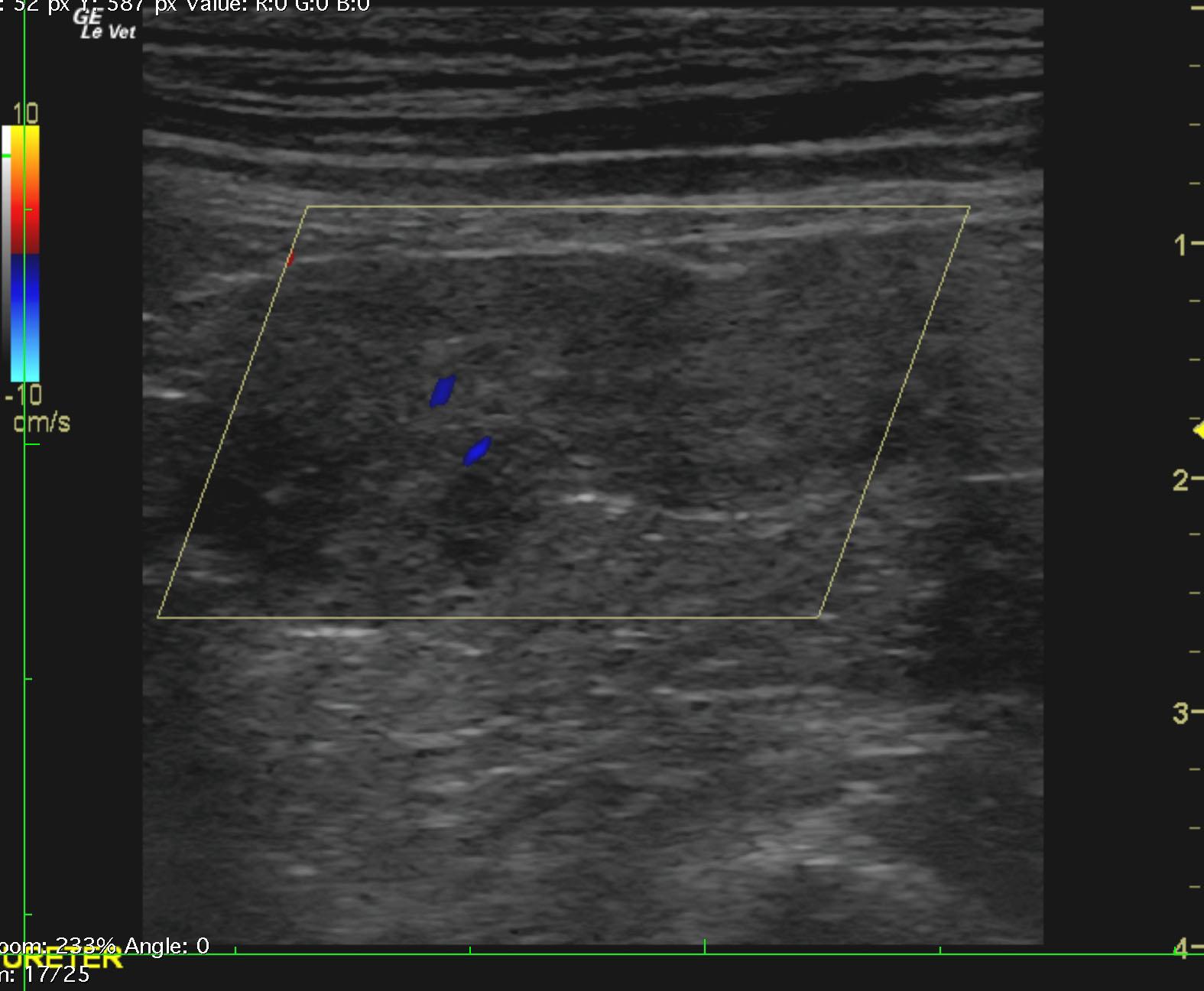
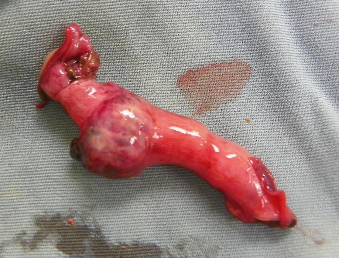
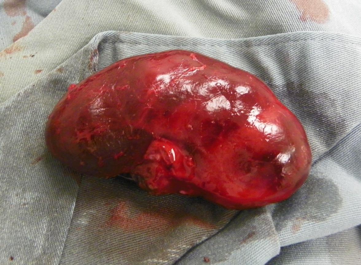
A right ureteral mass is visible obstructing urine flow from the right kidney with mass extrusion or blood clot present in the right kidney pelvis which is severely dilated as a result of hydronephrosis (Images 1-3). The lack of color flow in image 3 in the ureter is juxtaposed against the adjacent aorta that reveals a positive color flow signal. Image 4 shows a high resolution view of the ureteral mass, which is hypoechoic to surrounding fat. Color flow signals are present in the ureteral tissue owing to the neoplastic blood flow, which differentiates the mass from a clot or debris. The primary portion of the obstructive ureteral mass measures 3.53 x 1 cm (Image 5). Video clip of the ureteral mass shows evidence of neoplastic blood flow pattern (Video 1). High-resolution image of the obstructive ureteral mass demonstrates the obstructive position of the tumor with resultant hydronephrosis of the right kidney (Video 2-3). Video of US-guided FNA of the ureteral mass and post sampling view demonstrates no visible urine leakage or hemorrhage (Video 4-5). The potential for needle tracking of the tumor was discussed with the client as a potential risk should transitional cell carcinoma be the underlying cause. The right ureter/mass and hydronephrotic right kidney are shown after surgical resection (Images 6-7).
DX
Ureteral sarcoma
Outcome
The patient recovered uneventfully. Oncology consultation pending.
Comments
Case of the month from the sonographic “Zebra” category. If I ever put out another pathology DVD, I think this case would headline it, along with other obscure unexpected diagnoses by the probe: perhaps call it the ” The Zebra DVD of Sonographic Pathology.” Hematuria usually makes us think calculi, UTI or lower urinary tumors. But this one was a bit different. This was brought to us by my good friend, colleague, and sonopath specialist Dr. Doug Casey of English Bay US, Vancouver, BC, Canada (http://www.sonopath.com/specialists_casey.asp). Special thanks to Dr Phil Anderson and staff of the Animal Care Clinic (http://animalcareclinicvancouver.com) for the excellent management of this case.
Clinical Differential Diagnosis
Lower urinary tract disease – neoplasia, uroliths, bacterial cystitis, trauma. Prostatic disease – neoplasia, abscess. Upper urinary tract disease – neoplasia, renolith, pyelonephritis, trauma
Sampling
US-guided FNA of the ureteral mass revealed epithelial atypia. Surgical excisional biopsy of the right ureteral mass revealed ureteral sarcoma with hemorrhage. The right kidney demonstrated hydronephrosis, pelvic hemorrhage, parenchymal atrophy, fibrosis, and mild interstitial lymphoplasmacytic nephrosis.
Video
Patient Information
Patient Name :
Hunter Q
Gender :
Male, Neutered
Species :
Canine
Type of Imaging : Ultrasound
Status :
Complete
Liz Wuz Here :
Yes
Code :
06_00120
Clinical Signs
- Hematuria
Images
Clinical Signs
- Hematuria
Urinalysi
- Bacteria Present
- Blood Present