5 yo FS Rotttie X seen for 24 hr vomiting, inappetance, 1 wk hx of diarrhea in early April. No hx of dietary indiscretion.
- Hypokalemia, neutrophila & leukocytosis on B/W
- Painful abdomen on palpation with firm palpable structure mid abdomen
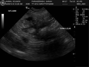
Scan revealed complex large mass like structure adjacent to ileo-colic-cecal junction, no obstrution or free air. No other significant findings.
5 yo FS Rotttie X seen for 24 hr vomiting, inappetance, 1 wk hx of diarrhea in early April. No hx of dietary indiscretion.
- Hypokalemia, neutrophila & leukocytosis on B/W
- Painful abdomen on palpation with firm palpable structure mid abdomen
Scan revealed complex large mass like structure adjacent to ileo-colic-cecal junction, no obstrution or free air. No other significant findings.
- In the first video for the orginal scan you can see the thickened terminal ielum dorsal and caudal (at 5:00 on the image) to the mass
- Second video show transverse of same area
The patient was rescanned 3 weeks later, was doing much better, eating but still had PM soft stool.
- Re-scan showed increase in mass size, slightly diminshed ileal thickness, closer to normal.
- FNA done later by rDVM had pyogranulmatous inflammation with reactive fibroplasia consistent with nodular steatitis, no neoplastic cells seen. Recommend biopsy for further evaluation.
- But dog is doing better!
So my question is this: has anyone seen anything like this and what would cause this bizarre configuration with no foreign body or neoplasia?

Comments
FNA is FNA and often when
FNA is FNA and often when neoplasia is surrounded by necrosis and inflammation and even tissue sepsis it is not diagnostic or misses the neoplasia especially in carcinoma. My tehcnique ont he fna of these is to stick each and every different echogenicity and pathological presentation so there is a big variety on the slide instead of sticking just one spot. theoretically helps:) This lesion meets neoplastic criteria and underlying lymphoma carcinoma or leio are all possible. On rare occasion spontaneous necrosis or non visible fb like a wood skewer piece will do this not be seen and smolder. Could core bx it or explore and aggressive resection. The intestine loses detail as well so lsa possible here or spontaneous intestinal necrosis. In these potential neoplasia with necorsis scenarios the patient is clinical for the inflammation and responds temporarily to med tx while the body gets things walled off. But as it continues to smolder it comes back or perfs. Carcinoma, lsa spontaneous necrosis/ibd or otehr neoplasia with inflammation/necrosis all possible. Resistant bacteria potential as well is possible.
Thanks Eric! I will forward
Thanks Eric! I will forward to rDVM, however dog is doing so much better the owner may opt to wait unfortunately.
Thanks Eric! I will forward
Thanks Eric! I will forward to rDVM, however dog is doing so much better the owner may opt to wait unfortunately.