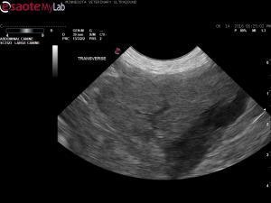
- 13 year old mn Rough Collie with polyuria-polydipsia and chronic diarrhea nonresponsive to budesonide
- Chem prof shows elevated ALKP, CBC shows a moderate anemia
- Abdominal US of the GI shows normal GI wall layering and thickness with no inflammation and no enlarged or reactive lymph nodes. The liver is also unremarkable.
- Within the splenic capsule, is a poorly defined, strongly hypoechoic lesion affecting the deep lateral aspect of the mid to caudal spleen. FNA’s were performed on the spleen and submitted to cytology.
- My differential diagnoses for the splenic lesion includes edema, hemorrhage due to trauma (dog has bilateral rear CP deficits), benign hemangioma, and neoplasia (MCT, hemangiosarcoma, other sarcoma). Coagulopathy has been ruled out based upon a normal PLT count (300,000) and normal PT/PTT.
- Any other thoughts on this splenic lesion? Should splenectomy be considered next if fna is nondiagnostic?
Thank you!
