Uterine mass in a 10 year old FI Standard poodle

Case Study

Uterine mass in a 10 year old FI Standard poodle

A 10-year-old intact female Standard poodle was presented for vomiting, which had not improved with symptomatic therapy. On abdominal palpation, a large firm abdominal mass was present.  
A 10-year-old intact female Standard poodle was presented for vomiting, which had not improved with symptomatic therapy. On abdominal palpation, a large firm abdominal mass was present.  
DX

Uterine mass

Sonographic Differential Diagnosis

Suspected uterine mass.

Image Interpretation

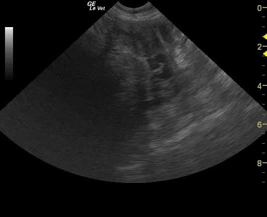
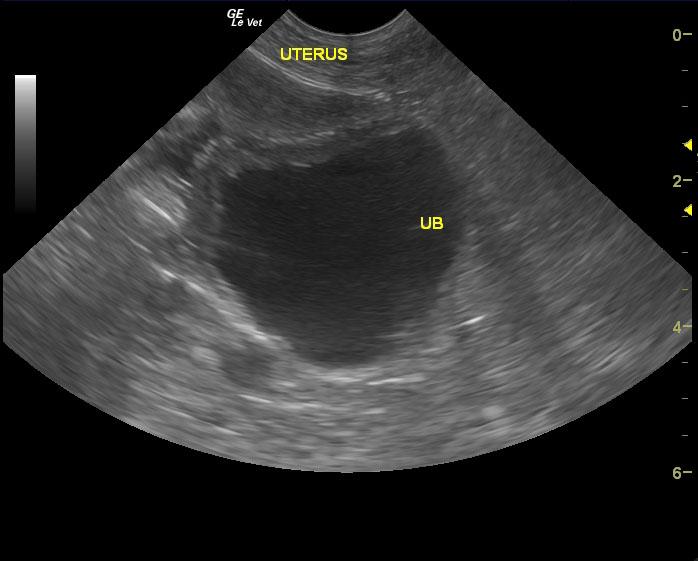
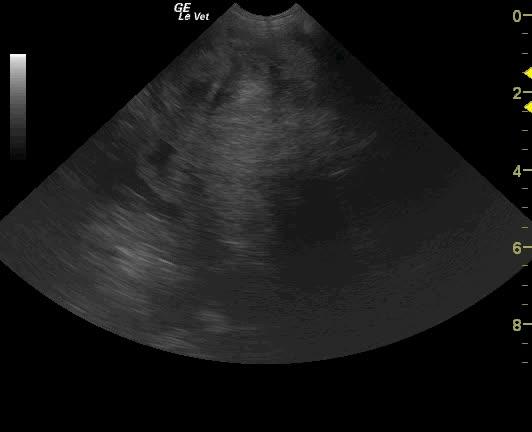
A fluid-filled body of the uterus can be visualized in the caudal abdomen. Tracing this fluid-filled uterus forward, a mixed echogenic mass was noted. Normal folds of the uterine endometrium can be visualized within the mass. This mass appears to be impinging on the spleen.

Outcome

Surgical exploratory surgery was performed and the mass was located in the uterine horn. No other disease was noted in the abdomen. The owners declined histopathology. The patient is currently doing well.

Clinical Differential Diagnosis

Palpable mass – neoplasia, granuloma, abscess, cyst of any organ system, hydronephrosis, pyometra

Sampling

None preoperatively. No histology available.

Patient Information

Patient Name : Wendy S
Gender : Female, Intact
Species : Canine
Type of Imaging : Ultrasound
Status : Complete
Liz Wuz Here : Yes
Code : 09_00059

Clinical Signs

  • Vomiting

Exam Finding

  • Palpable mass

Images

2012071022014000vlcsnap-2014-04-13-14h01m37s61vlcsnap-2014-04-13-14h02m18s148

Clinical Signs

  • Vomiting
Skip to content