Swollen kidneys and splenozaygos shunt in a 6yr old, FS, DSH.

Case Study

Swollen kidneys and splenozaygos shunt in a 6yr old, FS, DSH.

A 6-year-old FS DSH with a history of a long-term liver shunt that was maintained on occasional metronidazole and subcutaneous fluids was presented for evaluation of weight loss, hyporexia, and vomiting. CBC was within normal limits whereas serum biochemistry showed mildly elevated ALT activity, normal creatinine, and low urea.
A 6-year-old FS DSH with a history of a long-term liver shunt that was maintained on occasional metronidazole and subcutaneous fluids was presented for evaluation of weight loss, hyporexia, and vomiting. CBC was within normal limits whereas serum biochemistry showed mildly elevated ALT activity, normal creatinine, and low urea.

DX

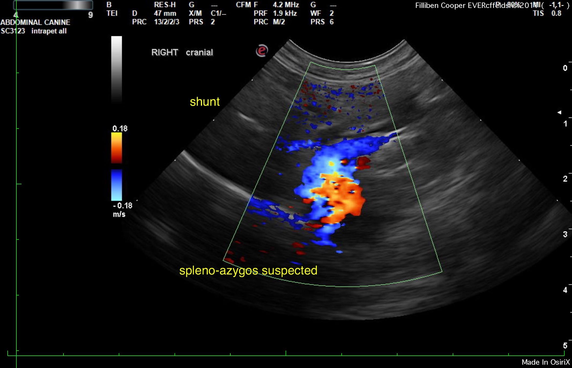
Swollen kidneys with medullary rim sign. Bladder sand. Anomalous extrahepatic portosystemic vessel consistent with splenoazygos shunt.

Sonographic Differential Diagnosis

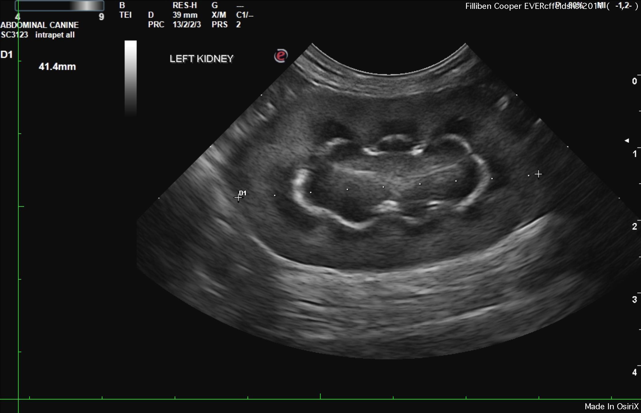
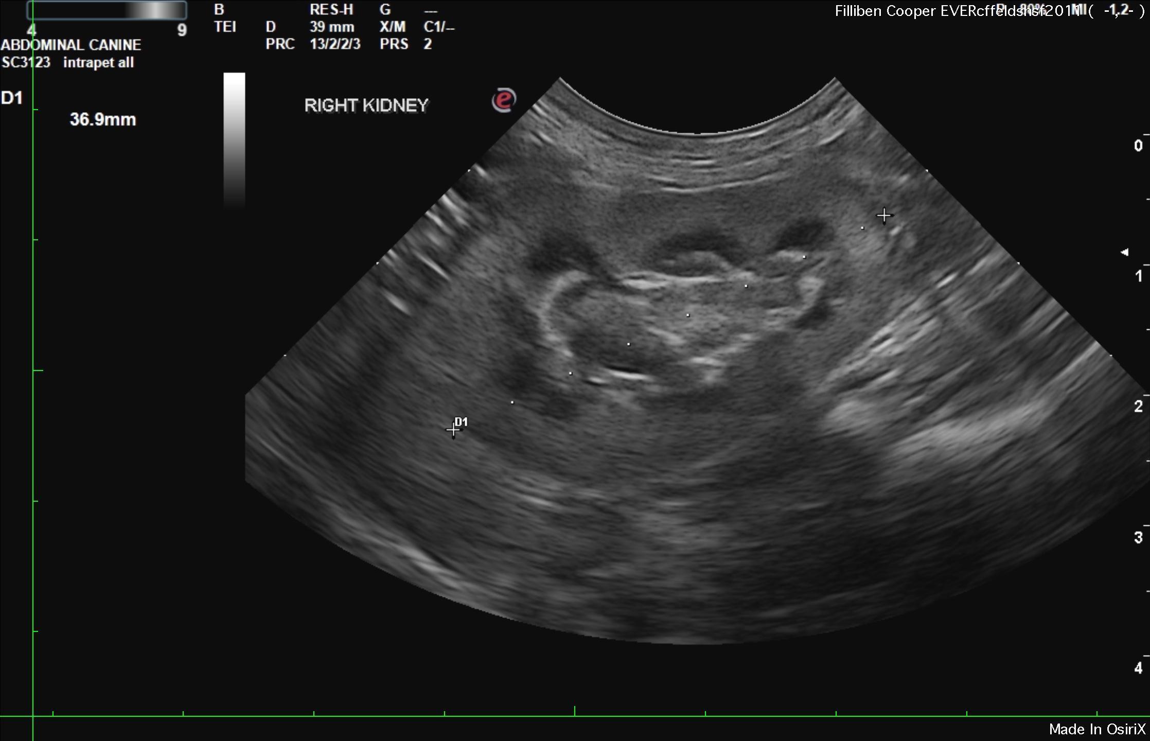
Swollen kidneys with medullary rim sign.
Bladder sand.
Anomalous extrahepatic portosystemic vessel consistent with splenoazygos shunt.

Image Interpretation

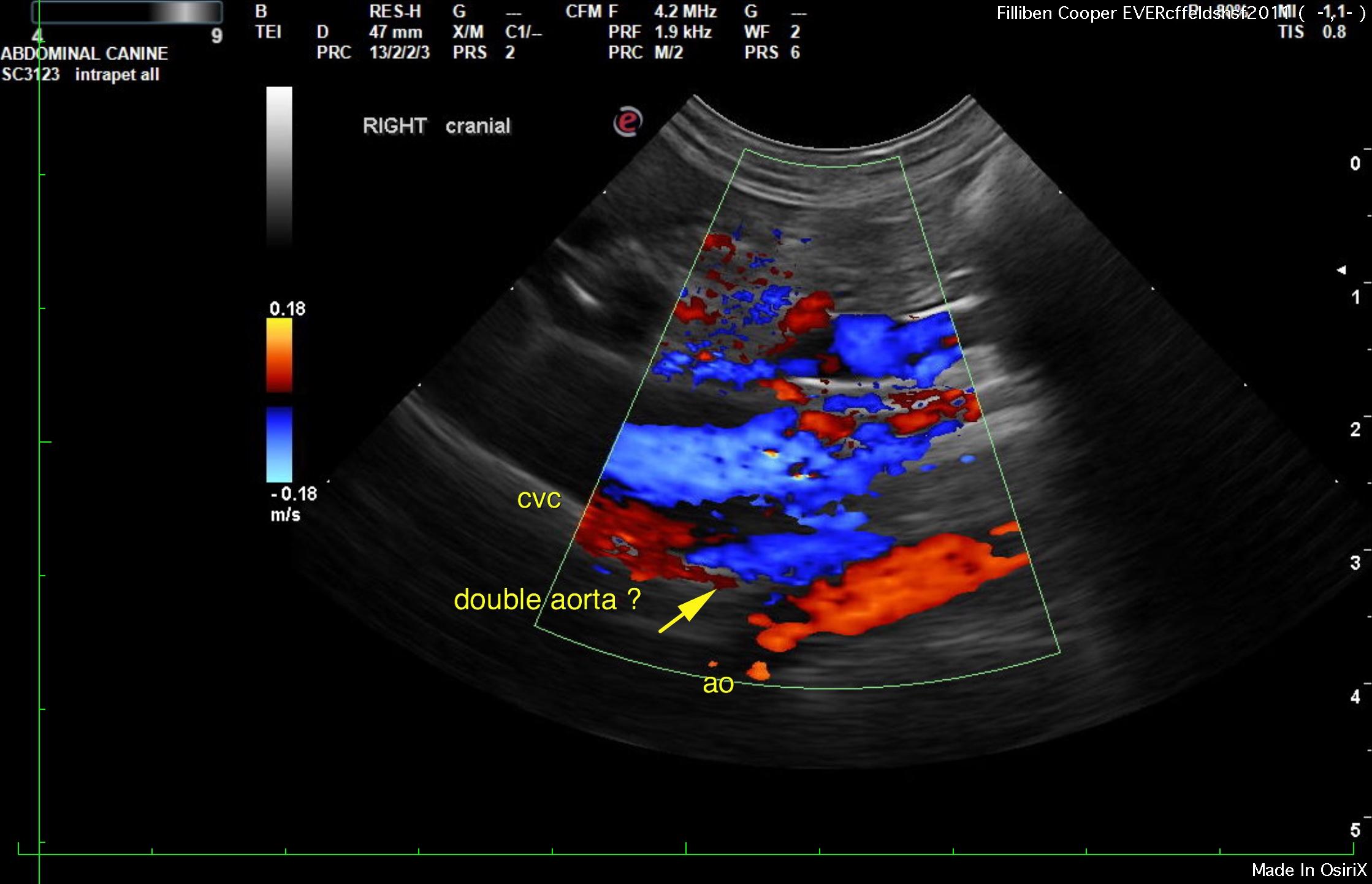
The liver was diffusely hyperechoic to the falciform fat. The vena cava and aorta ratio was 1:1. The vena cava measured 0.5 cm and the aorta also measured 0.5 cm. An anomalous vessel measuring approximately 0.8 cm in width decoursed dorsally and was deriving from the portal vein in the area of splenic vein derived extrahepatic portosystemic shunt. The vessel appeared to bypass the vena cava and enter into the diaphragm. This pattern is most consistent with splenoazygos shunt, yet ideally would be confirmed by CT evaluation. However, a dorsal connection to the vena cava could not be completely ruled out. One view demonstrated a third vessel, which would suggest a double aorta sign, which would be consistent with azygos shunt. The gallbladder and common bile duct were unremarkable.

The urinary bladder presented a minor amount of sand. 

The kidneys were swollen in this patient along with hyperechoic, medullary rim sign. The kidneys were hypervascular. The right kidney measured 3.69 cm. The left kidney measured 3.5 cm.

Outcome

Ideally CT evaluation would be performed for further definition and surgical planning. Bile acid profile is essential. Medical management may be adequate in this patient given the age. Typically splenoazygos are shunts that develop clinical signs later in life. Acute phased disease should be avoided at all costs in this patient such as gastrointestinal disease and pancreatitis. Strict dietary protocol is warranted.

Clinical Differential Diagnosis

Progression of liver shunt
Hepatopathy – cirrhosis, cholangio-hepatitis complex, neoplasia
Pancreas – pancreatitis, neoplasia

Patient Information

Patient Name : Copper Filliben
Gender : Female, Spayed
Species : Feline
Type of Imaging : Ultrasound
Liz Wuz Here : Yes

Clinical Signs

  • Anorexia
  • Vomiting
  • Weight loss

History

  • Portosystemic Shunt

Images

r1r2r4r9r10

Blood Chemistry

  • ALT (SGPT), High
  • BUN low

Clinical Signs

  • Anorexia
  • Vomiting
  • Weight loss
Skip to content