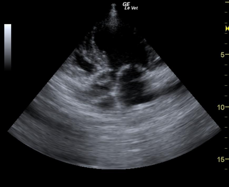
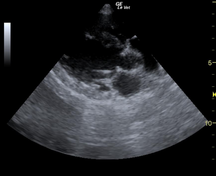
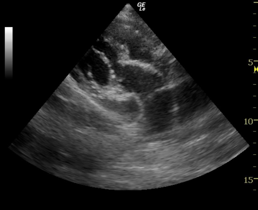
A 3-year-old neutered male Golden Retriever dog, with history of seizures and sub-aortic stenosis, presented for a re-evaluation to determine if the heart defect was stable and if any interventions would be appropriate at this time.
A 3-year-old neutered male Golden Retriever dog, with history of seizures and sub-aortic stenosis, presented for a re-evaluation to determine if the heart defect was stable and if any interventions would be appropriate at this time.