Splenic nodule in a 17 yr old, FS, DSH

Case Study

Splenic nodule in a 17 yr old, FS, DSH

A 17-year-old SF DSH was presented for evaluation of weight loss, reduced appetite, lethargy, and pain. Current therapy was methimazole and gabapentin. On survey radiographs, a ureterolith was evident.
A 17-year-old SF DSH was presented for evaluation of weight loss, reduced appetite, lethargy, and pain. Current therapy was methimazole and gabapentin. On survey radiographs, a ureterolith was evident.
DX

Splenic nodule

Sonographic Differential Diagnosis

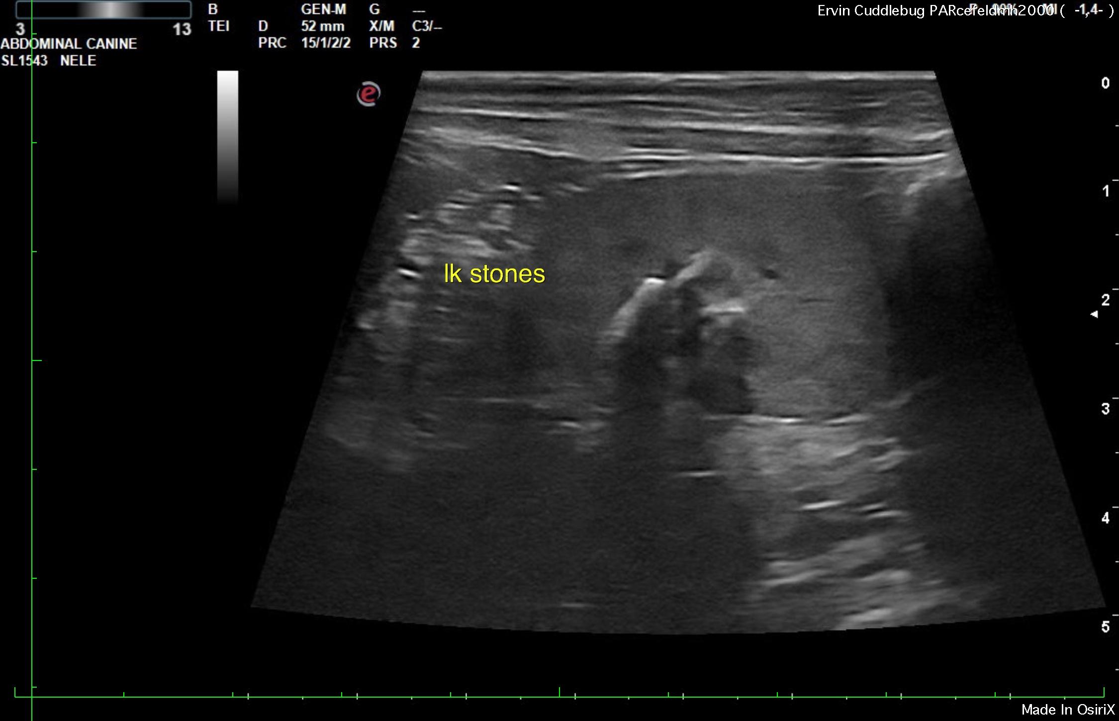
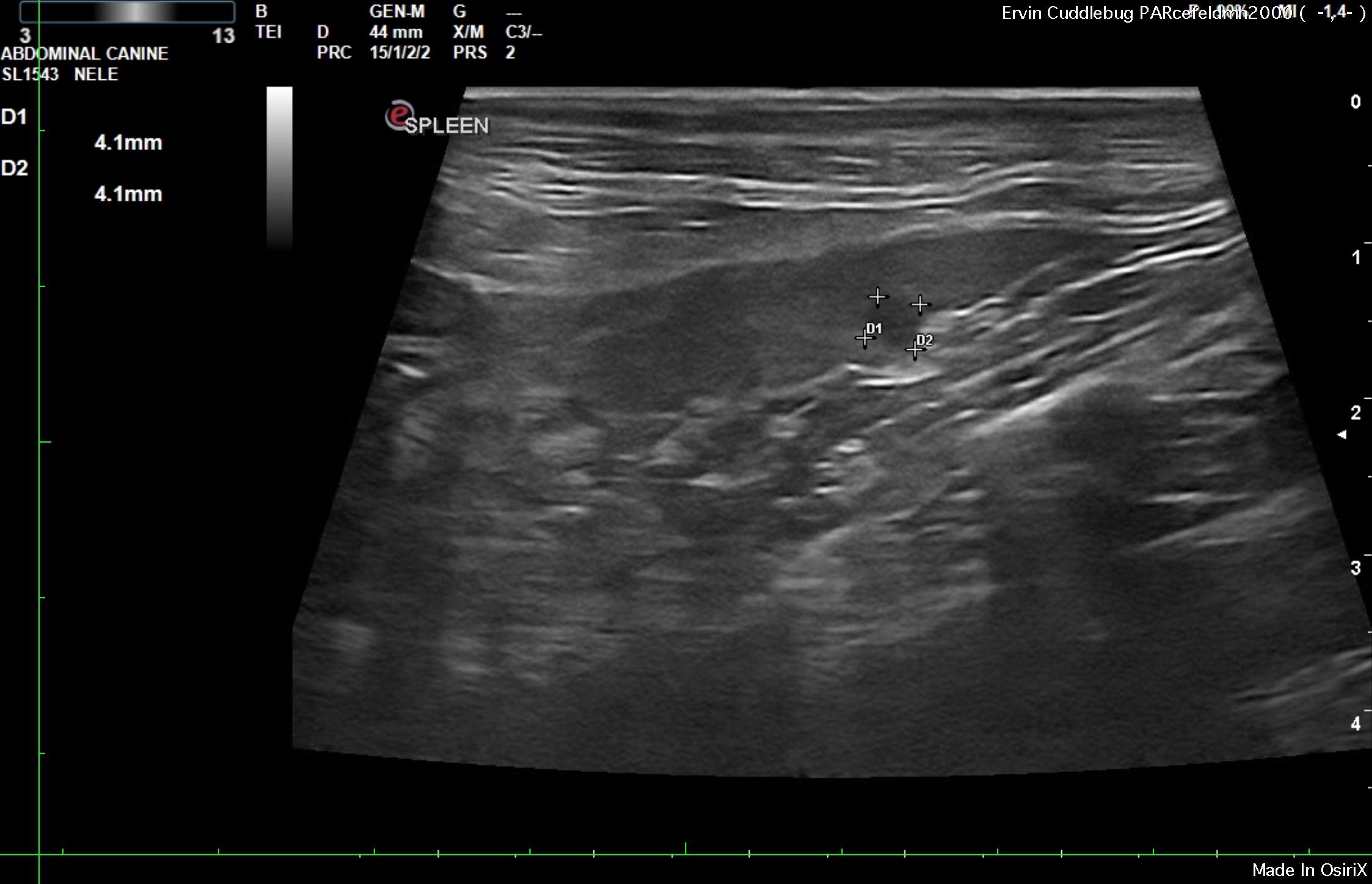
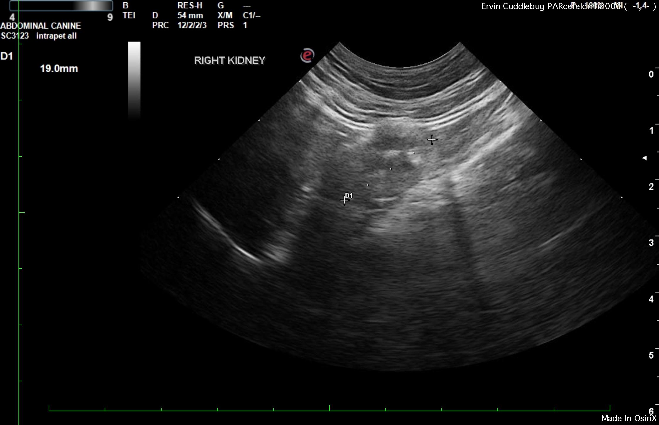
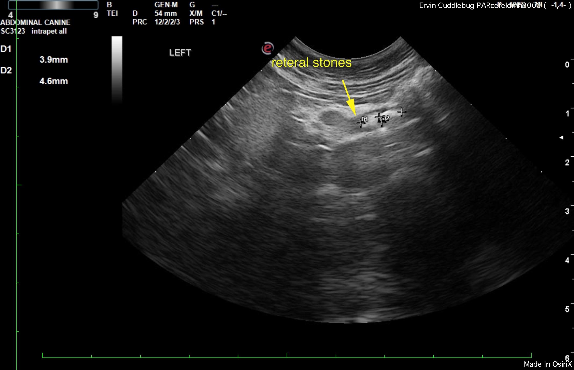
Splenic nodule. End stage renal dystrophy in the right kidney with ureteral calculi, slight pyelectasia and pelvic calculi in the left kidney with cortical infarcts, collapse and compensatory hypertrophy.
The renal pelvis in the left kidney in this patient is not dilated enough for potential ureteral bypass procedure with SUB device, as typically 0.5 cm of dilation is necessary for placement of SUB or stents. However, surgical intervention in order to liberate the ureteral stones would likely be in the patient’s best interest as the right kidney is likely not functional and the calculi appear to be embedded in the proximal ureter. Minor inflammatory/fibrotic pattern is noted around the left ureter. The calculi are obstructed approximately 2.5 cm caudal from the left renal pelvis. Nephrotomy to liberate the pelvic calculi may be in the patient’s best interest as well from a surgical standpoint as the largest calculus is nearly 1.0 cm. Other calculi are greater than 0.4-0.5 cm, which are typically too large to pass into the ureters. Perinephric inflammatory pattern was noted around the left kidney as well. Ideally the splenic nodule would be sampled.
Nodular hyperplasia is possible, yet relatively rare in cats. Abscessation or possible
round cell neoplasia are all possibilities.

Image Interpretation

The spleen revealed a focal, hypoechoic nodule that measured 0.41 cm with capsular expansion and disruption of architecture.

The kidneys revealed chronic interstitial nephrosis pattern from mild enlargement. The left kidney measured 4.5 cm with cortical infarcts and calculi with cortical collapse. Compensatory hypertrophy was noted in the cortices with hydroureter of 0.4 cm. The right kidney was severely dystrophic and measured 1.9 cm with fibrosis pattern, irregular contour, infarcts and calculi.

Clinical Differential Diagnosis

Ureterolith with obstructive uropathy
Renal – chronic kidney disease, neoplasia, renoliths, pyelonephritis

Patient Information

Patient Name : Cuddle Bug Ervin
Gender : Female, Spayed
Species : Feline
Type of Imaging : Ultrasound
Status : Complete
Liz Wuz Here : Yes

Clinical Signs

  • Anorexia
  • Lethargy
  • Pain
  • Weight loss

Images

a2a3a5a7

Clinical Signs

  • Anorexia
  • Lethargy
  • Pain
  • Weight loss
Skip to content