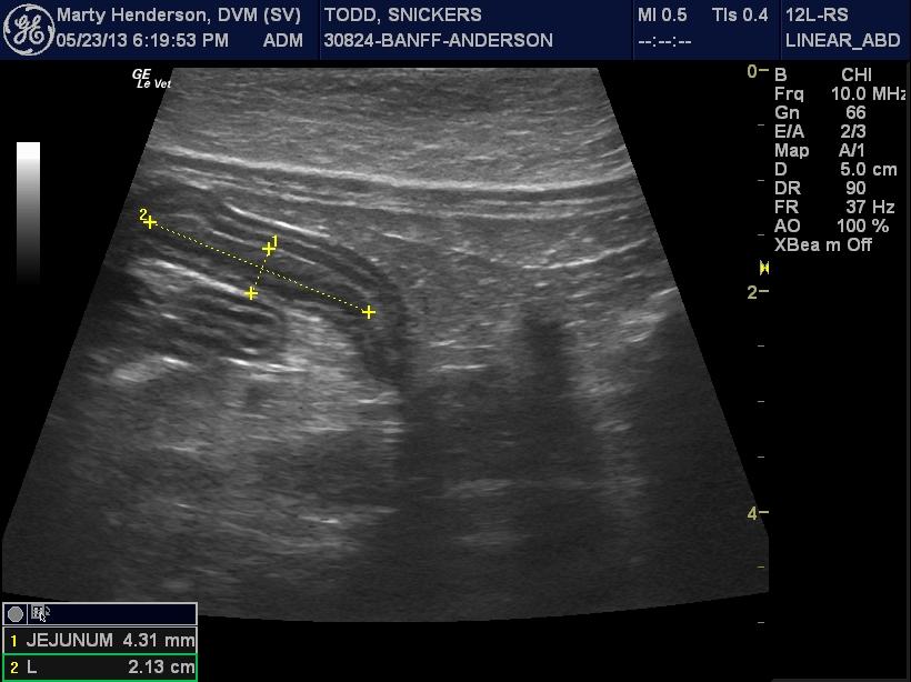
The patient is a feline DSH, NM, 10 years. The patient has intermittent vomiting and has lost 2 pounds in the past two months. Abdominal radiographs show slight accordion pattern.
The patient is a feline DSH, NM, 10 years. The patient has intermittent vomiting and has lost 2 pounds in the past two months. Abdominal radiographs show slight accordion pattern.